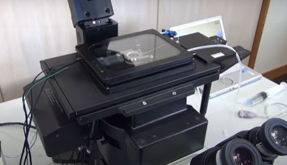
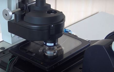
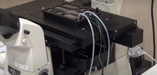
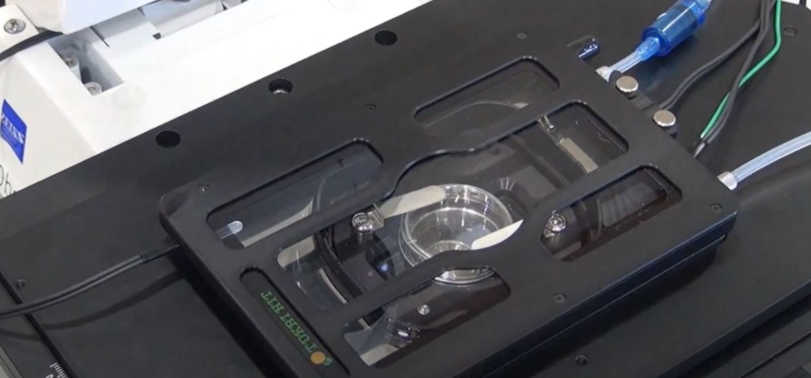
Live cell imaging을 위한 TOKAI HIT
Microscope Stage Top Incubator System
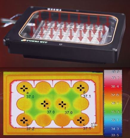
Microscope Time-Lapse Imaging을 위한 온도, 습도, CO₂조절시스템
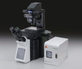
- Can be installed on general flat-top manual/motorized stage with 160 x 110 mm aperture.
- Optional Stage Adapter MK-IX3 (for IX3-SSU, IX3-SVR) and MK-SIG (for BX3-SSU, IX2-SFR/SVL2) are available.
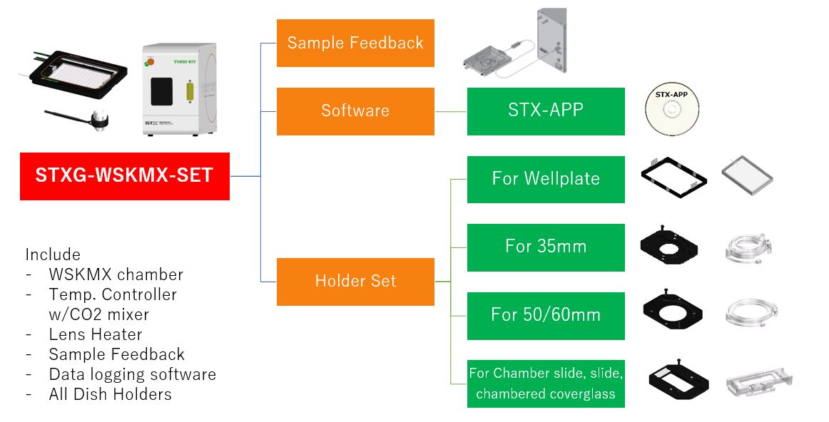
- Vessel holders are included in the package (except a holder for 35mm dish x 2pc).
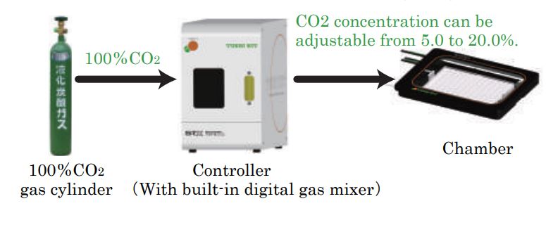
- Temp. Controller with built-in digital gas mixer for 100%CO2 gas use.
- CO2 concentration: 5.0 – 20.0%
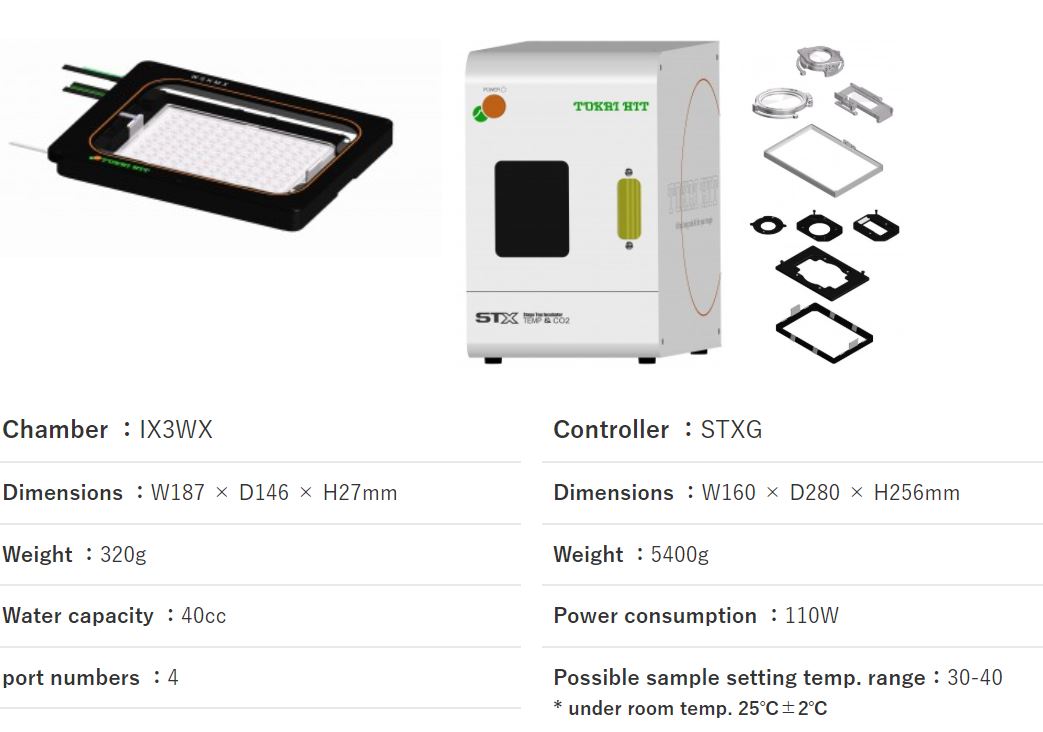
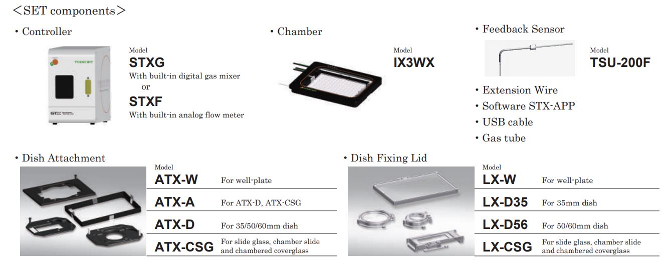
Support vessel / Dish Attachment
- Well:ATX-A
- 35mm:ATX-D
- 35mm×2:ATX-D35-2 (optional)
- 50mm:ATX-D
- 60mm:ATX-D
- Chamber slide :ATX-CSG
- Chambered cover glass :ATX-CGC
- Slide glass :ATX-CSG
Support Micrscope brand / Stage (required Adapter)
- OLYMPUS motorized: IX3-SSU[MK-IX3 (optional)]
- OLYMPUS motorized: BX3-SSU[MK-SIG (optional)]
- OLYMPUS manual: IX3-SVR[MK-IX3 (optional)]
- OLYMPUS manual: IX3-SVL[MK-IX3 (optional)]
- OLYMPUS manual: GX-SVR[MK-SIG (optional)]
- OLYMPUS manual: GX-SFR[MK-SIG (optional)]
- OLYMPUS manual: IX2-SFR/SVR[MK-SIG (optional)]

- Olympus IX83, Nikon Ti, Zeiss AxioObsever, Leica DMI4000B sut up
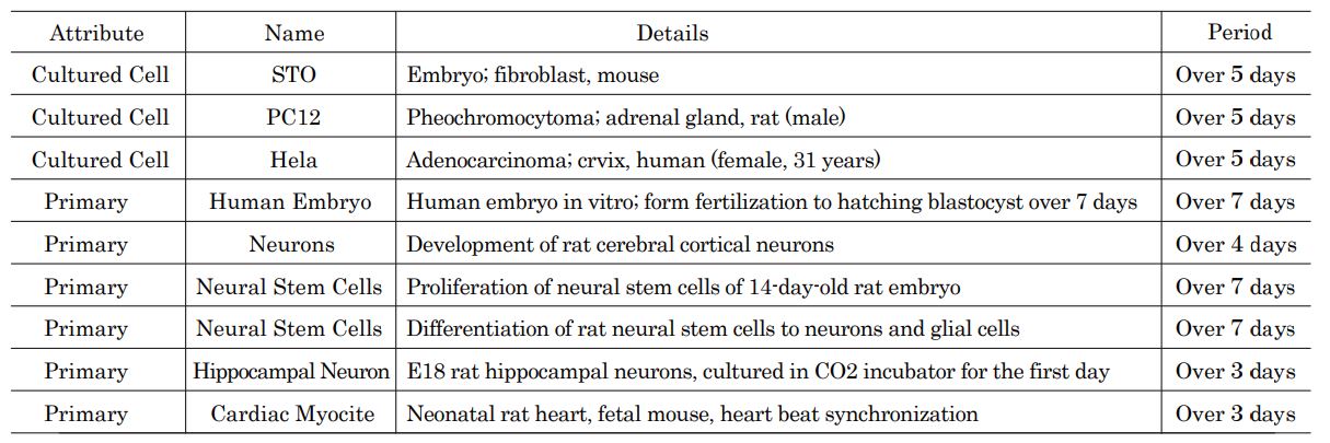
Applications
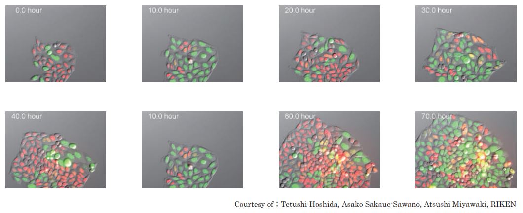
Courtesy of Dr. Simon Watkins, University of Pittsburgh Department of Cell Biology
By courtesy of Dr. Simon Watkins, University of Pittsburgh Department of Cell Biology
Courtesy of Moerner Lab, Stanford University
Courtesy of Lauren Piedmont Alvarenga of the Nikon Imaging Center @ Harvard Medical School
카다로그 다운로드

