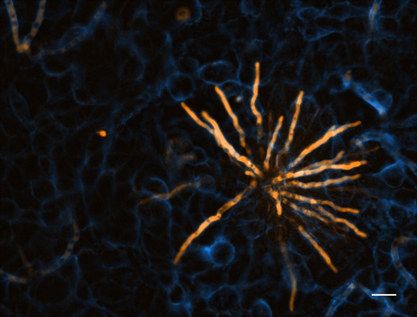
TdTomato-expressing Aspergillus fumigatus (amber) microcolony after 12 hours development on an A549 epithelial alveolar monolayer labelled with ConA-FITC (blue)
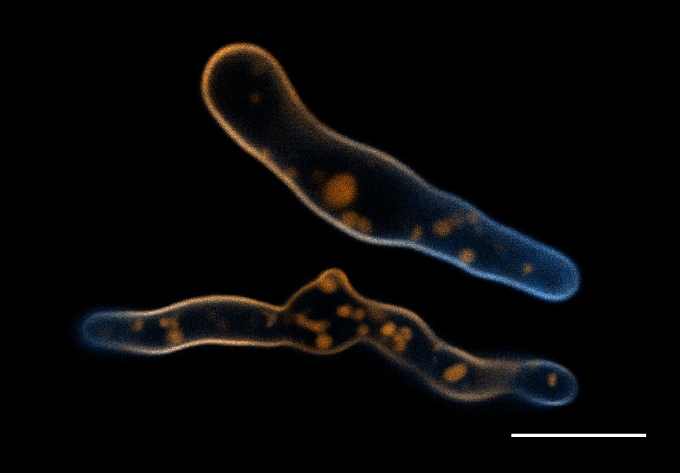
Aspergillus fumigatus germlings loaded with a synthetic cell membrane counterstain TMR-PAF96 (amber) and treated with fluorescent BODIPYcPAF26 anti-fungal peptide (cyan) to determine peptide’s localisation and mode of entry into the pathogen.
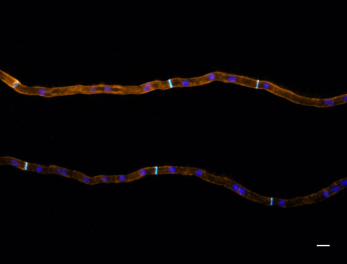
Neurospora crassa hyphal filaments treated with fluorescently labelled siRNA (amber) to determine its localisation, with respect to nuclei (blue) and the fungal cell wall and septa (cyan).
Ultrafast endocytosis at mouse hippocampal synapses taken with a CoolLED pE-2
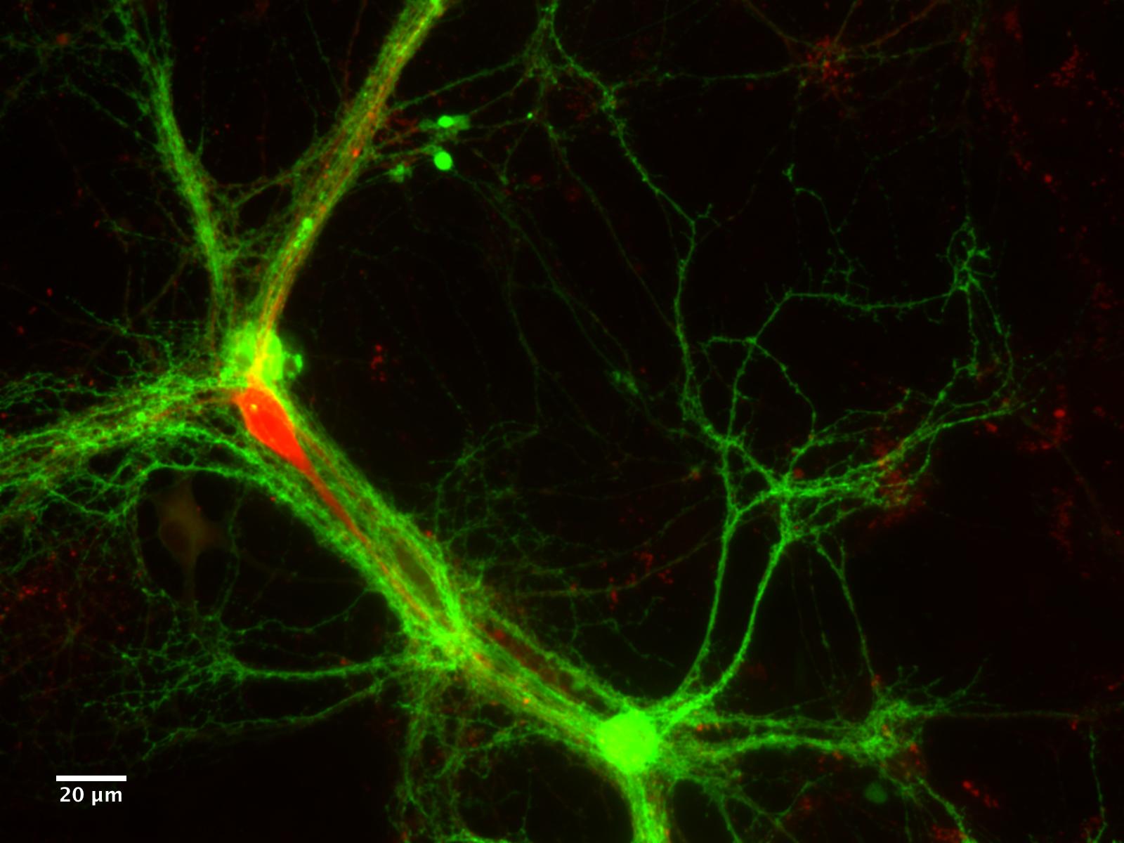
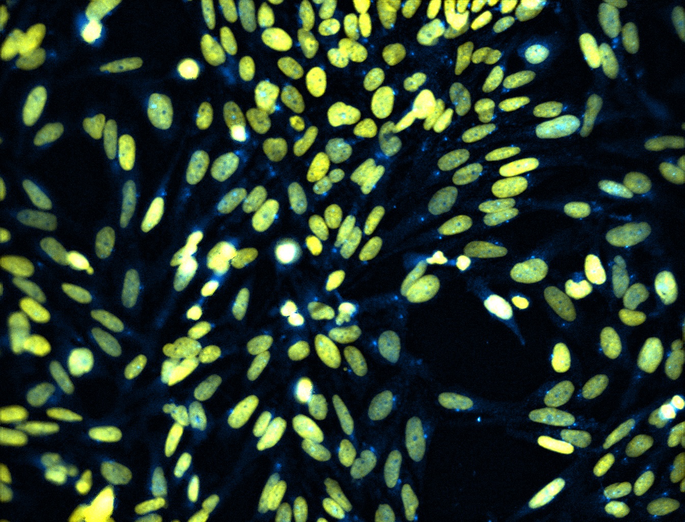
Antibody staining of a transcription factor localising in the nucleus
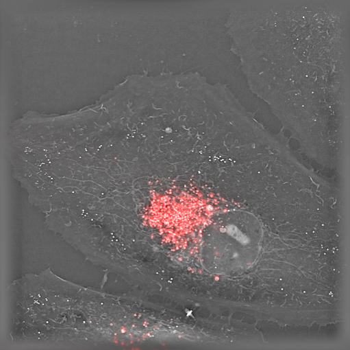
Live HeLa cells stained with lysosome marker imaged with Nanolive’s holotomographic technology.
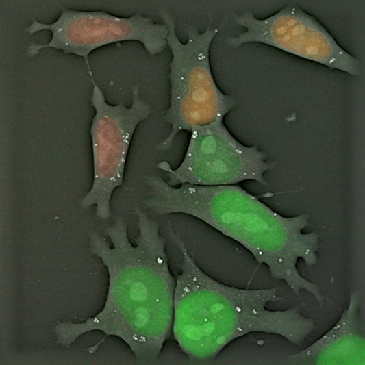
Live FUCCI mouse embryonic stem cells imaged with Nanolive’s holotomographic technology
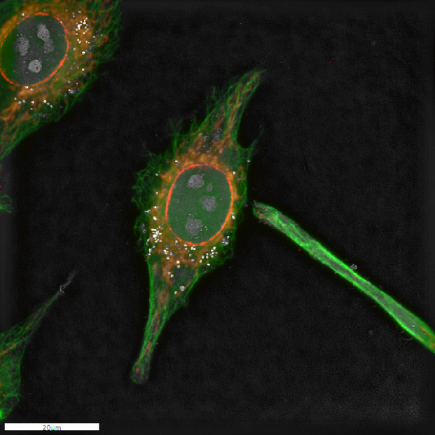
Fixed HeLa cells stained with Antibody for cytoskeleton (green), mitochondria (orange) and nuclear membrane(red). The whole cell was also imaged with Nanolive’s holotomographic technology and the four images were overlapped to create the overlay.
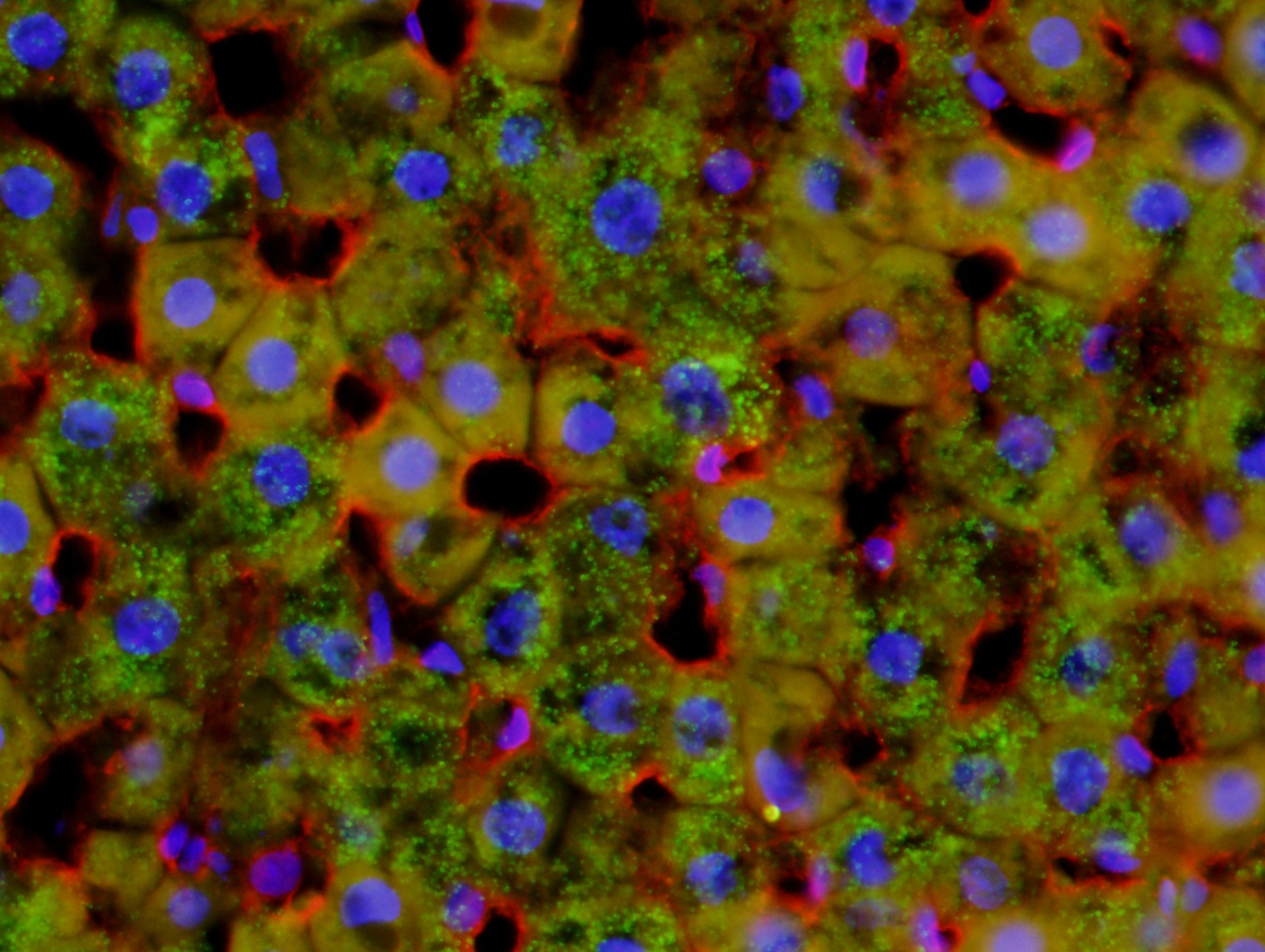
Fluorescent rat liver image taken using a CoolLED pE-300white, Olympus BX51 40x objective and a DP71 color camera
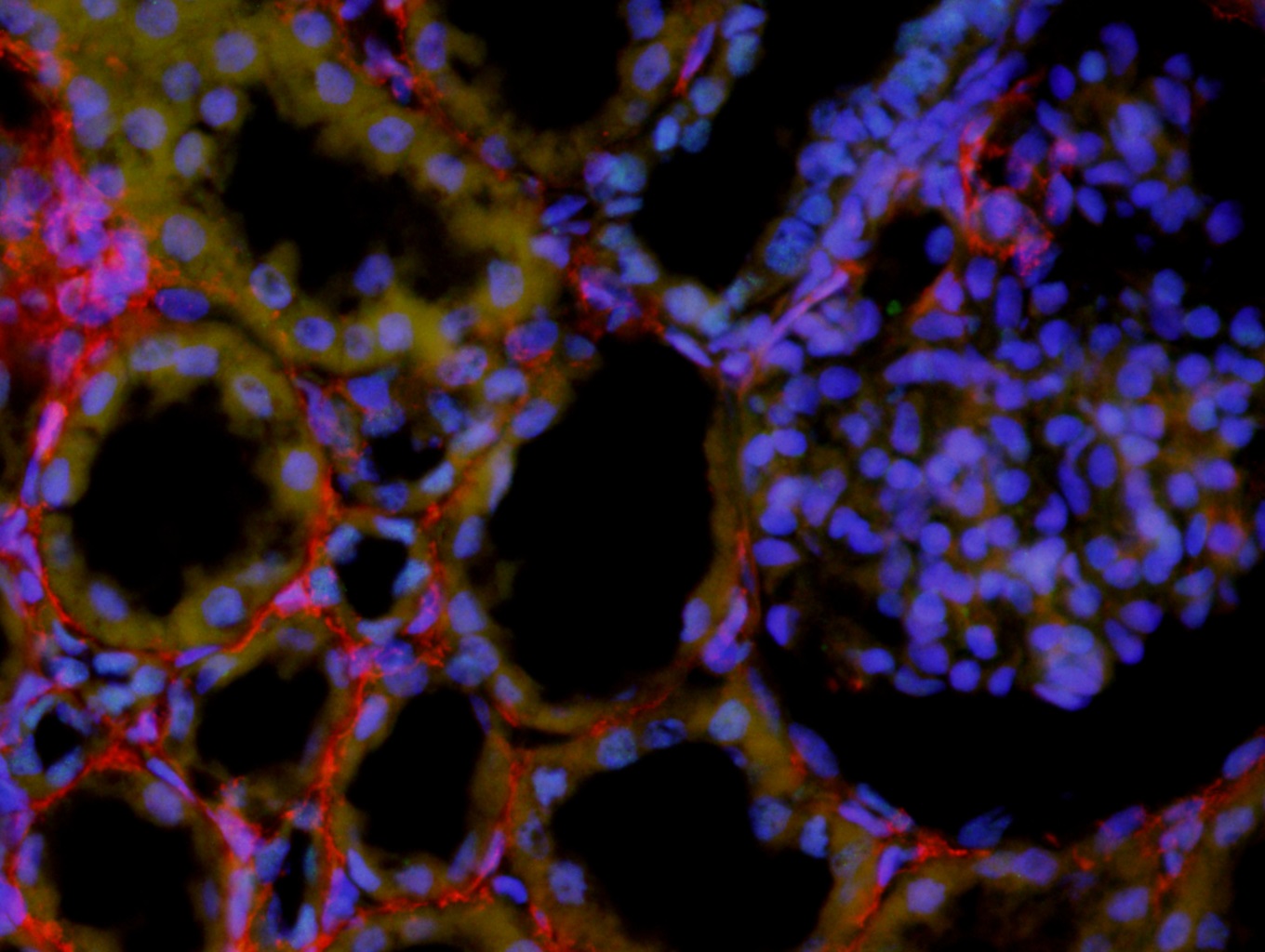
Fluorescent kidney image taken using a CoolLED pE-300white, Olympus BX51 40x objective and a DP71 color camera
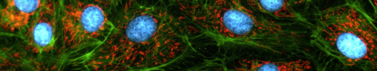
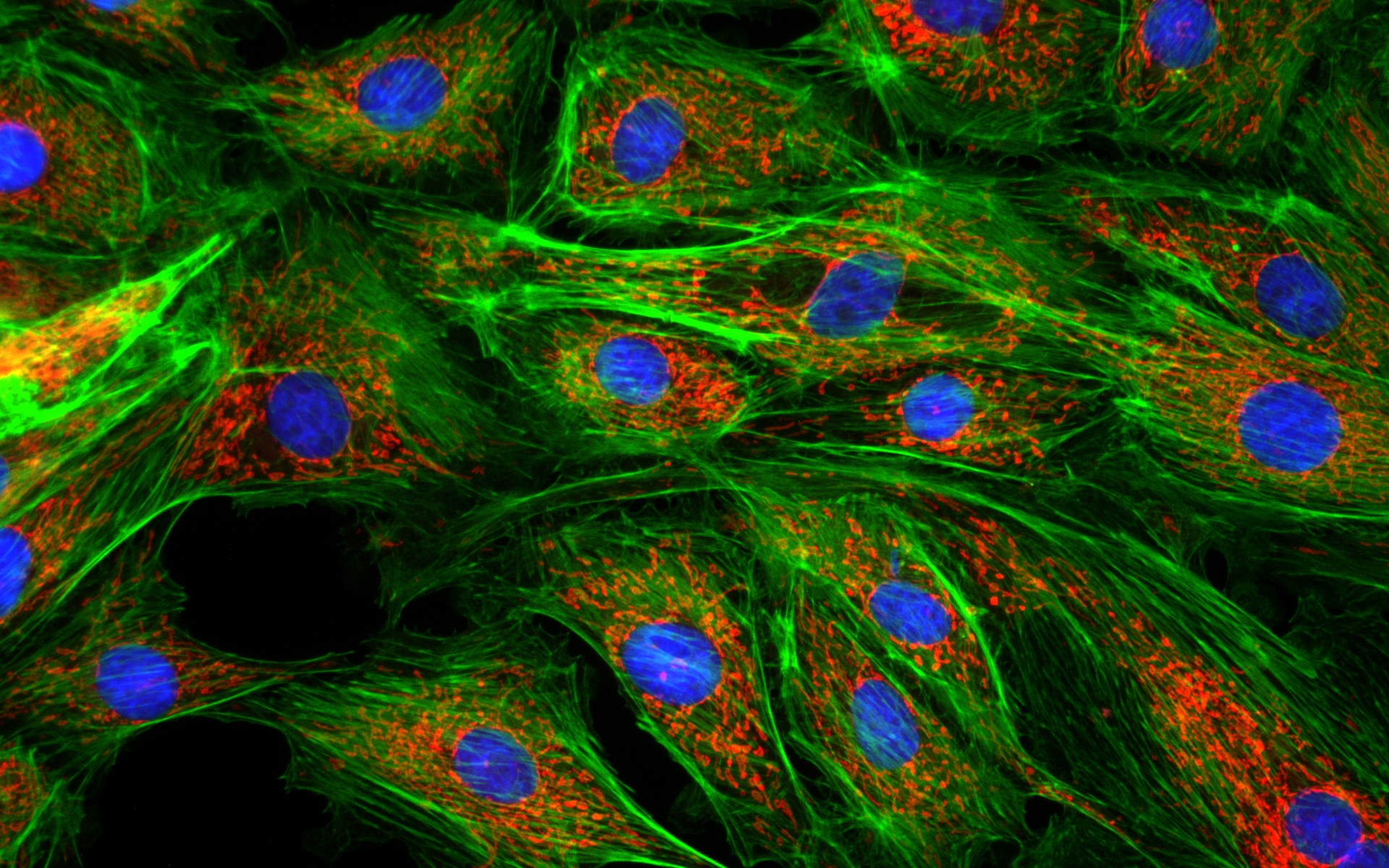
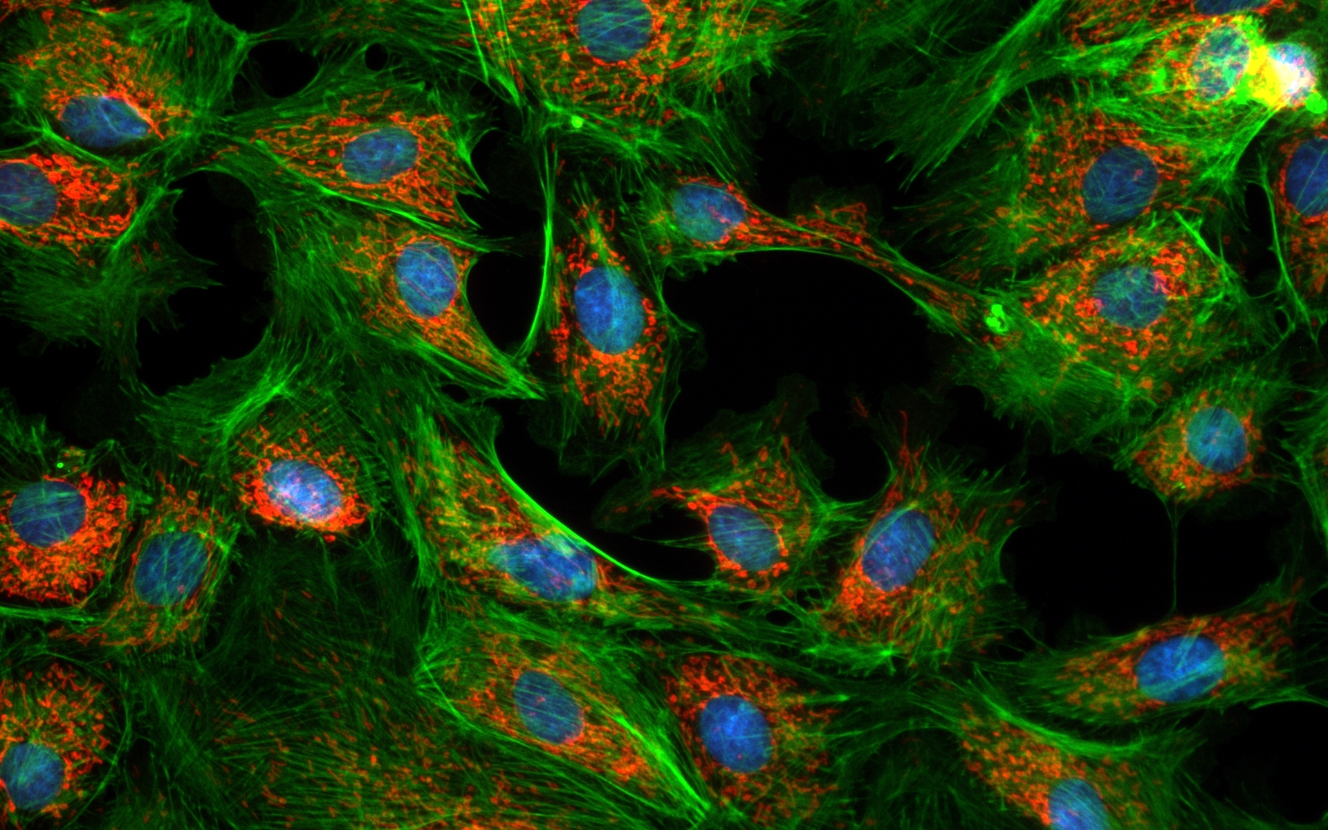
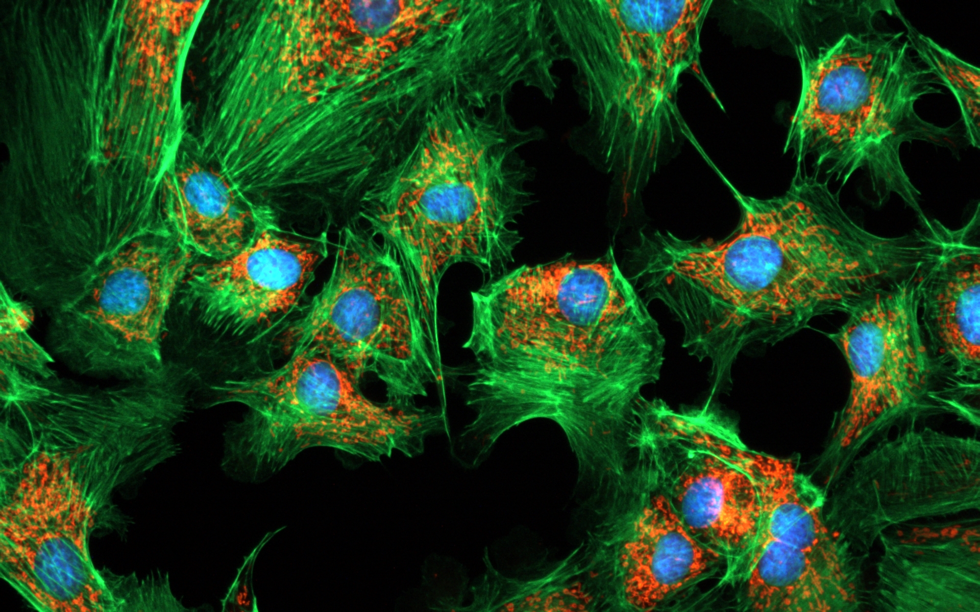
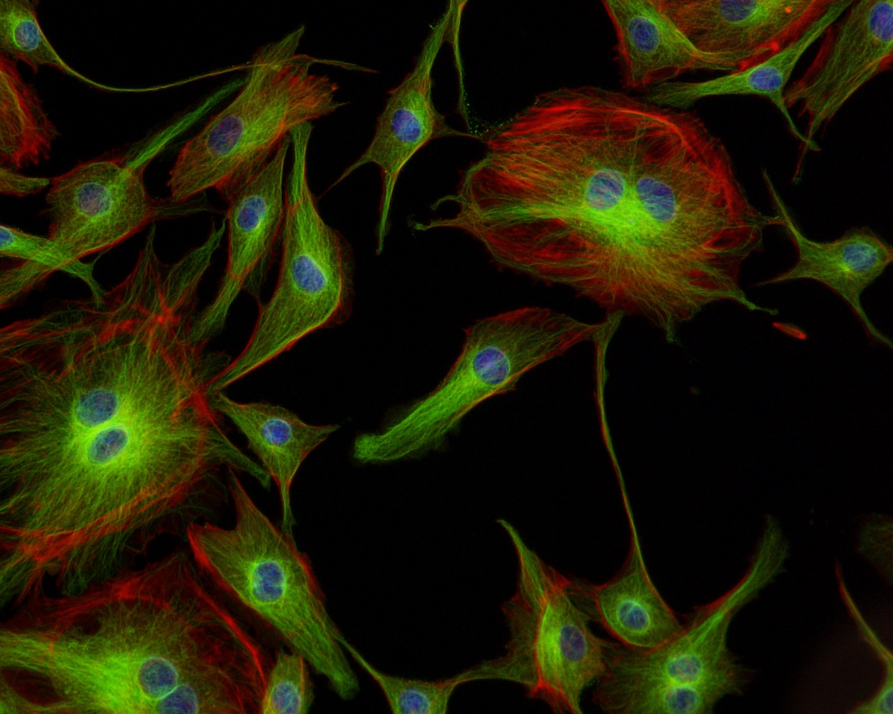
Fluorescent bovine pulmonary artery endothelial cells image taken using a CoolLED pE-300white, Nikon 20x objective and a black and white camera
Transmitted Podcarpus Elatus Leaf image taken using a CoolLED pE-100wht, Olympus BX51 20x objective and a DP71 color camera
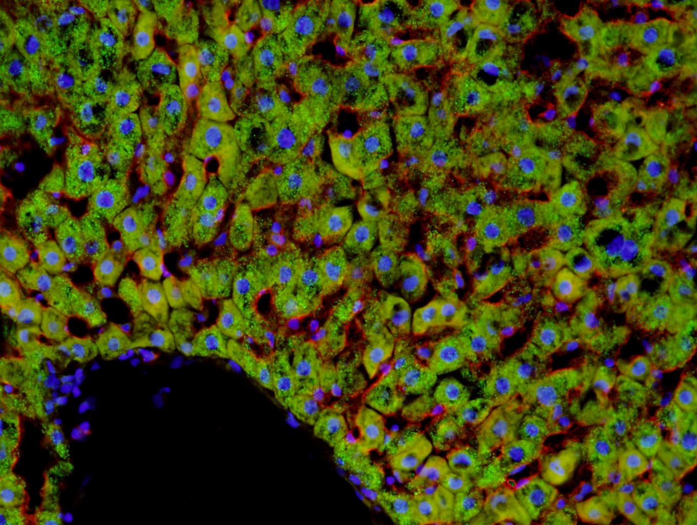
Fluorescent rat liver image taken using a CoolLED pE-300white, Olympus BX51 20x objective and a DP71 color camera
Fluorescent skin image taken using a CoolLED pE-300white, Olympus BX51 40x objective and a DP71 color camera