카테고리 보관물: Fluorescence

39007

JNOPTIC Filter 39007

Download Catalog_39007

39007 Spectrum

Filters Type
 AT620/50x EX
AT655DC BS
AT690/50m EM


– Filter set for Cy5/AlexaFluor 647/Draq5

  • Main Fluorochromes : Cy5
Excitation () Emission ()
649 670


Spectrum Comparison Fluor and 39007

Cy5 VS 39007

Best with these fluorochromes


39007 Spec

  • Typical Application(s) : Widefield Microscopy
  • Coating : Sputter/Hard Coated
  • Round excitation and emission filters mounted in anodized aluminum rings 2.3mm thick, up to 25mm in diameter
  • Rectangular dichroics up to 26x38mm, 1mm thick, to fit standard microscope manufacturer filter cubes


39010

JNOPTIC Filter 39010

Download Catalog_39010

39010 Spectrum

Filters Type
AT560/40x EX
AT600DC BS
AT635/60m EM


– Filter set for Texas Red/mCherry/AlexaFluor 594

Excitation () Emission ()
590 617


Spectrum Comparison Fluor and 39010

AlexaFluor 594 VS JNO-R1(B)


Best with these fluorochromes


39010 Spec

  • Typical Application(s) : Widefield Microscopy
  • Coating : Sputter/Hard Coated
  • Round excitation and emission filters mounted in anodized aluminum rings 2.3mm thick, up to 25mm in diameter
  • Rectangular dichroics up to 26x38mm, 1mm thick, to fit standard microscope manufacturer filter cubes

39004

JNOPTIC Filter 39004

Download Catalog_39004

39004 Spectrum

Filters Type
AT540/25x EX
AT565DC BS
AT605/55m EM


– Filter set for TRITC/Cy3/TagRFP/AlexaFluor546

  • Main Fluorochromes : TRITC
Excitation () Emission ()
555 580

Spectrum Comparison Fluor and 39004

TRITC VS 39004

Best with these fluorochromes


39004 Spec

  • Typical Application(s) : Widefield Microscopy
  • Coating : Sputter/Hard Coated
  • Round excitation and emission filters mounted in anodized aluminum rings 2.3mm thick, up to 25mm in diameter
  • Rectangular dichroics up to 26x38mm, 1mm thick, to fit standard microscope manufacturer filter cubes


39002

JNOPTIC Filter 39002

Download Catalog_39002

39002 Spectrum

Filters Type
AT480/30x EX
 AT505DC BS
AT535/40m EM


– Filter set for EGFP/FITC/Cy2/AlexaFluor488

  • Main Fluorochromes : FITC
Excitation () Emission ()
490 525

Spectrum Comparison Fluor and 39002

FITC VS 39002


Best with these fluorochromes


39002 Spec

  • Typical Application(s) : Widefield Microscopy
  • Coating : Sputter/Hard Coated
  • Round excitation and emission filters mounted in anodized aluminum rings 2.3mm thick, up to 25mm in diameter
  • Rectangular dichroics up to 26x38mm, 1mm thick, to fit standard microscope manufacturer filter cubes


39000

JNOPTIC Filter 39000

Download Catalog_39000

39000 Spectrum

Filters Type
 AT375/28x EX
AT415DC BS
 AT460/50m EM


– Filter set for DAPI/Hoechst/AlexaFluor350

  • Main Fluorochromes : DAPI
Excitation () Emission ()
359 461

Spectrum Comparison Fluor and 39000

DAPI VS 39000


Best with these fluorochromes


39000 Spec

  • Typical Application(s) : Widefield Microscopy
  • Coating : Sputter/Hard Coated
  • Round excitation and emission filters mounted in anodized aluminum rings 2.3mm thick, up to 25mm in diameter
  • Rectangular dichroics up to 26x38mm, 1mm thick, to fit standard microscope manufacturer filter cubes

49003

JNOPTIC Filter 49003

Download Catalog_49003

49003 Spectrum

Filters Type
ET500/20x EX
 T515LP BS
ET535/30m EM


– Filter set for EYFP

  • Main Fluorochromes : YFP
Excitation () Emission ()
514 527


Spectrum Comparison Fluor and 49003

YFP VS 49003


Best with these fluorochromes


49003 Spec

  • Typical Application(s) : Widefield Microscopy
  • Coating : Sputter/Hard Coated
  • Round excitation and emission filters mounted in anodized aluminum rings 2.3mm thick, up to 25mm in diameter
  • Rectangular dichroics up to 26x38mm, 1mm thick, to fit standard microscope manufacturer filter cubes

mCherry

Spectrum Chart

mCherry Spectrum
Excitation () Emission ()
587 610


Recommended Filter set

  • 일반 형광 Lighting source 사용시

Filter Model No. Excitation filter (㎚) Dichroic mirror (㎚) Emission filter (㎚) Remarks
JNO-R1(B) 560/40
(540~580)
600 (LP) 635/60
(605~665)
Wide Excitation band,
Bandpass Emission
49008 560/40
(540~580)
600 (LP) 630/75
(592.5~667.5)
Wide Excitation band,
Bandpass Emission
49306 580/25
(567.5~592.5)
600 (LP) 625/30
(610~640)
Narrow Excitation band,
Bandpass Emission


  • 레이저 Lighting source 사용시

Filter Model No. Excitation filter (㎚) Dichroic mirror (㎚) Emission filter (㎚) Remarks
49910 561/10
(556~565)
561 (LP) 630/70
(595~665)
For use with laser between 557~563

Narrow Excitation band,
Bandpass Emission

49911 594/10
(589~599)
594 (LP) 645/75
(607.5~682.5)
For use with laser between 591~596

Narrow Excitation band,
Bandpass Emission

Information

mCherry is a fluorophore (a fluorescent protein) used as a marker when tagged to molecules and cell components. mCherry and the majority of red fluorescent proteins derive from the protein drFP583 (commonly known as DsRed) isolated from Discosoma sea anemones, while other fluorescent proteins in the green range are often variants of jellyfish GFP. DsRed is a tetramer optimally excited at 558 and has an emission maximum at 583 .

mCherry는 형광 물질 (형광 단백질)로서 분자 및 세포 구성 요소에 태그를 부착 할 때 marker 로 사용됩니다. mCherry와 대다수의 적색 형광 단백질은 Discosoma 말미잘에서 분리된 단백질 drFP583 (일반적으로 DsRed라고 함)에서 유래한 반면, 녹색 범위의 다른 형광 단백질은 종종 해파리 GFP의 변종입니다. DsRed는 558 ㎚에서 최적으로 여기되고, 583 ㎚ 에서 최대 방출을 갖는 4 량체입니다. 

EBFP

Spectrum Chart
BFP Spectrum
Excitation () Emission ()
385 448
Recommended Filter set
  • 일반 형광 Lighting source 사용시

Filter Model No. Excitation filter (㎚) Dichroic mirror (㎚) Emission filter (㎚) Remarks
49021 405/20
(395~415)
425 (LP) 460/50
(435~485)
Narrow Excitation band,
Bandpass Emission
49028 395/25
(382.5~407.5)
425 (LP) 460/50
(435~485)
Narrow Excitation band,
Bandpass Emission


  • 레이저 Lighting source 사용시

Filter Model No. Excitation filter (㎚) Dichroic mirror (㎚) Emission filter (㎚) Remarks
49901 405/20
(395~415)
405 (LP) 460/50
(435~485)
For use with laser between 400~410

Narrow Excitation band,
Bandpass Emission

Information

Fluorescent proteins, like EBFP, can be used as protein “tags” to study the subcellular localization of proteins and/or their translocation upon stimulation or as markers for transfection in transient and stable expression systems.

EBFP와 같은 형광 단백질은 자극이나 전이를 위한 안정적인 marker 로서 또는 그들의 일시적인 발현의 하위 세포를 연구하기 위해 단백질 “태그” 로 사용될 수 있습니다.


ACQUCAM 23GR IMAGES

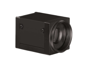
AcquCAM 23GR


More images with AcquCAM 23GR


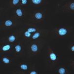
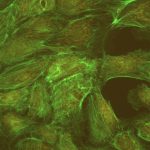
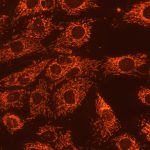
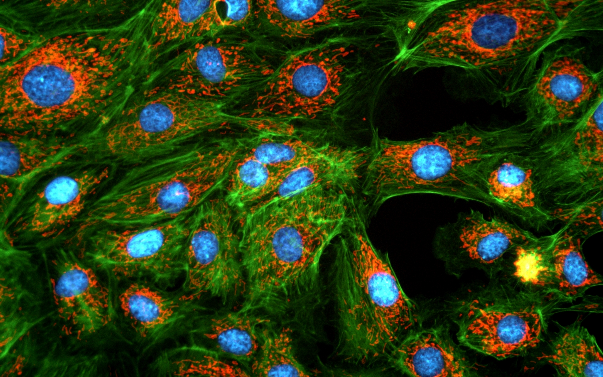
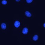
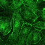
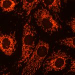
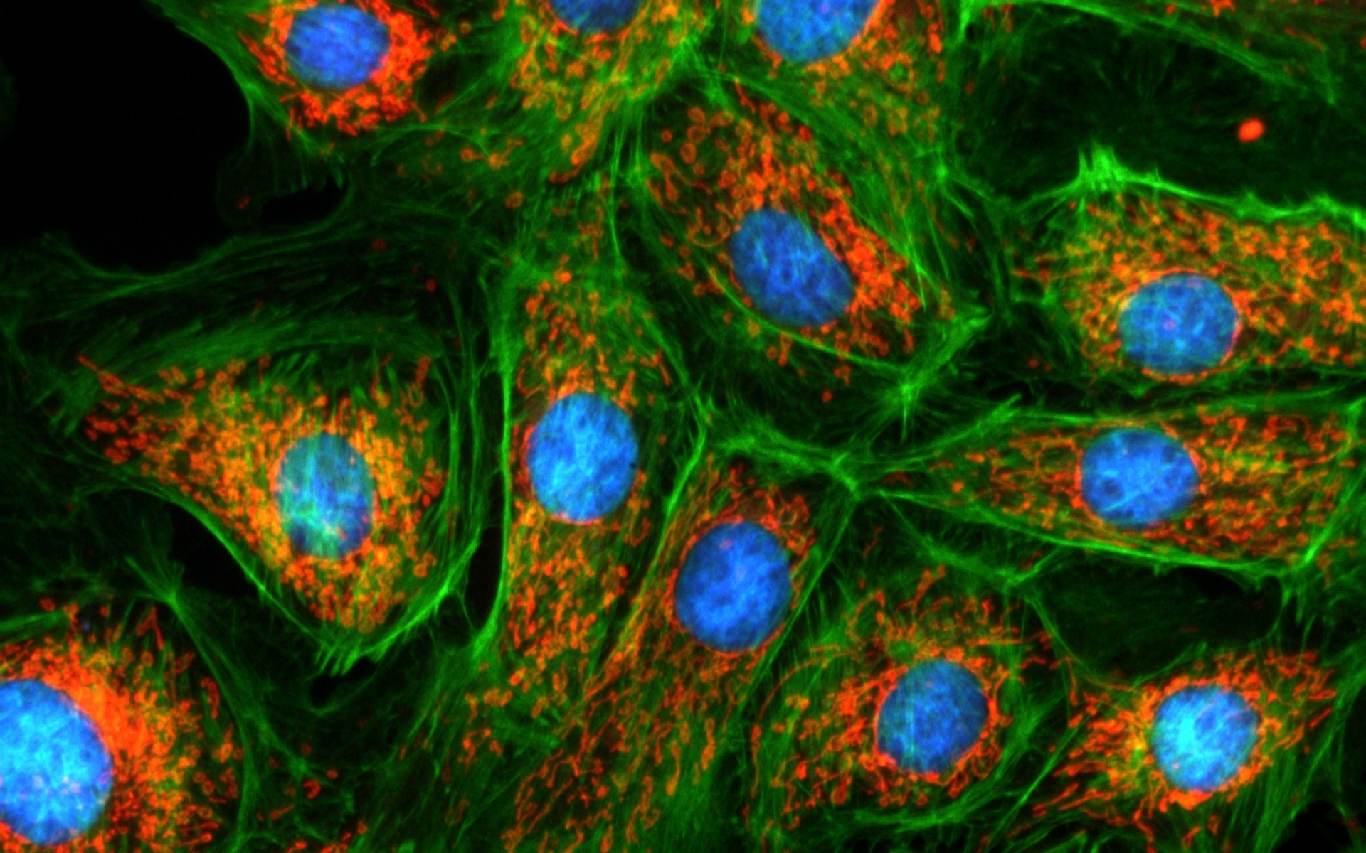
Sample : bovine pulmonary artery endothelial cell, BPAEC
Filter Set : JNO-U(L), B(L), G(L)
Camera : AcquCAM 23GR2
C-mount adapter : 1X
Lens : UPLNAPO40X
Above three images are merged and processed using ARM S/W


Sample : bovine pulmonary artery endothelial cell, BPAEC
Filter Set : JNO-U(B), B(B), G(B)
Camera : AcquCAM 23GR2
C-mount adapter : 1X
Lens : PlanApo60xWLSM
Above three images are merged and processed using ARM S/W


AcquCAM 23GR

AcquCAM 23GR


(Down load Catalog  – ACQUCAM 23GR)


  • USB 3.0 Color Camera
  • 1/1.2 ” Exmor Sensor
  • 1920×1200 pixel
  • Trigger input and I/O
  • Available  of taking images with a wavelength of 650nm or more, such as CY5
  • High Sensitivity Sensor (Best)
  • Fast, seamless image transfer
  • Available of images with a very wide field of view
  • Low Noise Sensor


The displays live digital images with gradual smoothness and combines exceptional resolution  with faithful color reproduction.The new sensors feature a global shutter function and able to capture a high-speed moving image without focal plane distortion. High-speed processing, low noise and low power dissipation by using column-parallel A/D conversion. equipped with trigger mode, and the external pulse can control accumulation time. The Sensor also have a pulse output function to indicate respective conditions during shutter operation and can be coordinated with peripheral circuits. High-definition images can be displayed live at a rate of high frames per second. without compression. Such imaging quality enables even the finest cellular regions to be observed clearly and distinctly without deterioration. While focusing is made stress free.

Images with AcquCAM 23GR


Sample : bovine pulmonary artery endothelial cell, BPAEC
Filter Set : JNO-U(B), B(B), G(B)
Camera : AcquCAM 23GR2
C-mount adapter : 1X
Lens : UPLNAPO40X
Above three images are merged and processed using ARM S/W


JNO-ARM

Advance Realtime Monitor. Easy-to-Use, Image Enhancement, etc


1. Information

ARM is image analysis software for JNOPTIC AcquCAM cameras. This S/W is interchangeable with all WDM cameras regardless of camera brand and model and even more the most strength point is simple and easy use of length, area, angle, etc.


2. Simple measurement tools

  • Measurement Tools: Count, Distance, Angle, Area, etc.
  • Guide Lines: Cross Line, Rectangular Lattice, Circular Grid
  • Scale Panel on Live/Image Screen
  • Available measurement on LIVE image and saved image mode


3-1. Specialization for observation of fluorescence images

Improvement Function about Fluorescence Microscopy Image. (Used function: Auto Level, Low level, Pseudo Color, Merge Image)

Bottom on the left is merged image without improvement of image. And the bottom on the right is merged image after improvement of image. These images are shot by JNOPTIC AcquCAM Pro/G3 camera


3-2-1 Effect of high-contrast image with simple operation (Monochrome)

Improvement Function about Microscopy Image Monochrome. (Used function: Auto Level)

Effective improvement of image with simple operation. This image is shot by JNOPTIC Pro/S3 camera


3-2-2 Effect of high-contrast image with simple operation (Color)

Improvement Function about Microscopy Image. (Used function: Auto Level)

Effective improvement of image with simple operation. These images are shot by JNOPTIC AcquCAM Pro/G3 camera


3-3-1 Live Pseudo Color (for color camera)

When you get fluorescence images using color camera, if the unwanted channel was shown because of cross talk, you could remove this channel to take advantage of function of Live Pseudo.
Improvement Function about Fluorescence Microscopy Live Camera Image. (Used function: Live Pseudo Color)
 The extraction of wanted color information from color image (Remove BR on RGB source) These images are shot by JNOPTIC AcquCAM Pro/G3 camera

3-3-2 Live Pseudo Color (for Monochrome camera)

When you get fluorescence images using monochrome camera, you can raise efficiency of acquisition for fluorescence images to add similar false color to the color to be shown on the eyepiece in real time.
Improvement Function about Fluorescence Microscopy Live Monochrome Camera Image. (Used function: Live Pseudo Color)
 Improvement of usage environment for monochrome camera to add false color on the image. These images are shot by JNOPTIC AcquCAM Pro/S3 camera

4. JNO-MHU(Option Unit)

Available measuring height with additional unit, JNO-MHU

※ JNO-MHU is sold separately as optional unit.

Left: Focus on top of sample (Z-axis reset), Right: Focus on bottom of sample (Z-axis height measurement)

 < Measuring condition : over 20x objective, temp 20°C >

Responding Model Measurement value unit Measurable height Error of measuring 1000㎛ Recommended measuring height
CX 41 0.2 ㎛ -9.9 ~ 29.9㎜ Below ±20㎛ Below ± 2000㎛
CKX 41 0.2 ㎛ -9.9 ~ 29.9㎜ Below ±20㎛ Below ± 2000㎛
BX – FM 0.2 ㎛ -9.9 ~ 29.9㎜ Below ±20㎛ Below ± 2000㎛
BX 51/53 0.1 ㎛ -9.9 ~ 29.9㎜ Below ±10㎛ Below ± 1000㎛
MX 51 0.1 ㎛ -9.9 ~ 29.9㎜ Below ±10㎛ Below ± 1000㎛
MX 61L/61 0.1 ㎛ -9.9 ~ 29.9㎜ Below ±10㎛ Below ± 1000㎛


5. Reporting to Excel


Specification

Spec Description
Size 1/1.2 inch
Resolution H: 1920, V: 1200
Pixel size H: 5.86 µm, V: 5.86 µm
Video formats& Frame rate 1920×1200 RGB24@ 54 fps
1920×1200 Y800@ 162 fps
1920×1200 Y16@ 80 fps                             1920×1200 Y411 @ 108 fps
1920×1200 YUY2 @ 81 fps
Dynamic range 8 / 12 bit
Exposure time 20 ㎲ to 30s
Lens mount C/Cs mount
Interface USB 3.0
Power supply 4.5 to 5.5 VDC
Dimensions H: 29 mm, W: 29 mm, L: 43 mm
Mass 65g


More images with AcquCAM 23GR

-> For more details, click here.