Jae Hwan Jin의 모든 글

VF-200-0Z Vibratome

 CompresstomeⓇ VF-200-0Z 의 특징 : 

  • 엔트리급 Compresstome®
  • 전기 생리학 및 이미징 연구를 위한 생체 조직을 얇게 슬라이스하는 데 사용되는 반자동 조직 슬라이서
  • 특허의 슬라이스 기술을 통해 건강한 티슈의 생존 능력을 두 배로 높임
  • 사용자 친화적인 고속의 슬라이싱 작업

VF-200-0Z 의 적용 가능 분야 :

  • 전기생리학
  • 기관형배양
  • 폐의 정밀 절단용

VF-200 의 Technical Spec :

Technical Specification VF-200-0Z
Advance Speed 1-20 ㎜/s, adjustable
Return Speed 20 ㎜/s, fixed
Vibration Frequency 0-20 ㎐, adjustable
Vibration Amplitude 2 ㎜, fixed
Z-axis Vibration ~0 ㎛
Blade double edge stainless,
ceramic blades
Cutting Angle 13 degrees,
fixed(standard)
Thickness Adjustment manual
Micrometer Resolution 10 ㎛/div
Maximum Tissue
Diameter
12.5 ㎜ (standard tube),
15.5 ㎜ (large tube),
6 ㎜ (small tube)
Maximum Tissue
Length
25 ㎜
Minimum Slice
Thickness
10 ㎛
Cutting Mode single
Bath 140 x 60 x 30 ㎜
Power Source DC 13-15 V
Power Consumption 500 mW
Dimension ( L x W x H ) 360 x 210 x 190 ㎜
Weight 5 ㎏

Down load  Technical Specification_VF-200-0Z

VF-200-0Z 부품 : 

VF-200-0Z  소모품 : 


Compresstome® VF-200-0Z vibrating microtome

The Compresstome®  VF-200-0Z is our entry level Compresstome®. With our patented compression technology, this semi-automated slicer excels at sectioning slices used for:
Our patented slicer technology allows you to double your slice viability. The Compresstome® is also easy to use, and our online training videos will allow everyone in your lab to produce excellent slices.

The Compresstome VF-200-0Z is best used to section live tissue for electrophysiology and imaging studies. If you are looking to section fixed tissue for immunohistochemistry, please explore the VF-300-0Z.

With our patented compression technology, this semi-automated slicer excels at sectioning slices used for:

Products we recommend to purchase with VF-200-0Z:

To reduce contamination when working with both fresh and fixed tissue we recommend purchasing additional:

Consumables we recommend for quality use include:


Down load  Technical Specification_VF-200-0Z


VF-300 Vibratome

CompresstomeⓇ VF-300 의 특징 : 

  • Compresstome® 제품군의 주력 제품
  • 완벽하게 자동화된 슬라이스 기술
  • 특허의 슬라이스 기술을 통해 건강한 티슈의 생존 능력을 두 배로 높임
  • 편리한 사용

VF-300 의 적용 가능 분야 :

  • 전기생리학
  • 기관형배양
  • 폐의 정밀 절단용

VF-300 의 Technical Spec :

Technical Specification VF-300
Advance Speed 0-20 ㎜/s, adjustable
Return Speed 20 ㎜/s, fixed
Vibration Frequency 0-20 ㎐, adjustable
Vibration Amplitude 2 ㎜, fixed
Z-axis Vibration < 2 ㎛
Blade double edge stainless,
ceramic blades
Cutting Angle 13 degrees,
fixed(standard)
Thickness Adjustment digital adjustment,
automatic
Micrometer Resolution 1 ㎛/step
Maximum Tissue
Diameter
15.5 ㎜ ((standard tube),
6 ㎜, 12.5mm (small tube)
Maximum Tissue
Length
25 ㎜
Minimum Slice
Thickness
4 ㎛
Cutting Mode single/ continuous selectable
Bath 140 x 60 x 30 ㎜
Power Source DC 13-15 V
Power Consumption 4 W
Dimension ( L x W x H ) 330 x 240 x 190 ㎜
Weight 7 ㎏
Down load  Technical Specification_VF-300


Training  Videos :

VF-300 Embedding & Slicing
VF-300 Maintenance
VF-300 Changing & aligning Blade
The VF-300 is the workhorse of the Compresstome®family.  With our compression technology, this fully automated slicer excels at sectioning slices used for:
Our slicer technology allows you to double your slice viability. The Compresstome®  is also easy to use. Our online training videos will allow everyone in your lab to produce excellent slices.

At Precisionary Instruments, we aim to push the boundaries of scientific research. That’s why we love to see how scientists find new ways to use our products.

At the Allen Institute, the “open science” concept pairs neuroscientists with system engineers to come up with innovative ideas and conjure them into existence. We are proud that the Compresstome® can be part of this project, helping the scientists work with both mouse brains as well as human brain tissues.

Products we recommend to purchase with VF-300-0Z:

To reduce contamination when working with both fresh and fixed tissue we recommend purchasing additional:

Consumables we recommend for quality use include:

Down load  Technical Specification_VF-300

VF-300-0Z Vibratome
 CompresstomeⓇ VF-300-0Z 의 특징 : 
  • 최신의 vibratome 기술의 진보
  • 특허받은 압축 기술을 통해 경쟁사보다 우위를 확보
  • 아가로 오스(Agarose) 삽입 과정에서 부드럽고 균일한 압축은 섹셔닝 중에 티슈를 안정화
  • 부드럽고 건강한 티슈 제작
  • Auto Zero-Z® 기술로 직접 Z 축 튜닝을 할 필요가 없음

VF-300-0Z 의 적용 가능 분야 :
  • 전기생리학
  • 면역조직 염색
  • 기관형배양
  • 폐의 정밀 절단용

VF-300 의 Technical Spec :

Technical Specification VF-300-0Z
Advance Speed 0-20 ㎜/s, adjustable
Return Speed 20 ㎜/s, fixed
Vibration Frequency 0-20 ㎐, adjustable
Vibration Amplitude 2 ㎜, fixed
Z-axis Vibration ~0 ㎛
Blade double edge stainless,
ceramic blades
Cutting Angle 13 degrees,
fixed(standard)
Thickness Adjustment digital adjustment,
automatic
Micrometer Resolution 1 ㎛/step
Maximum Tissue
Diameter
15.5 ㎜ (standard tube),
6 ㎜, 12.5mm (small tube)
Maximum Tissue
Length
25 ㎜
Minimum Slice
Thickness
4 ㎛
Cutting Mode single/ continuous selectable
Bath 140 x 60 x 30 ㎜
Power Source DC 13-15 V
Power Consumption 4 W
Dimension ( L x W x H ) 330 x 240 x 190 ㎜
Weight 7 ㎏

Down load  Technical Specification_VF-300-0Z
VF-300-0Z 부품 : 

VF-300-0Z  소모품 : 

The VF-300-0Z Compresstome® from Precisionary Instruments represents the latest advances in vibratome technology. Our patented compression technology enables our vibratome to overpower the competition. This gentle, uniform compression from the agarose embedding process helps to stabilize the tissue during sectioning and allows our vibratome to produce smoother and healthier slices than the competition. The VF-300-0Z is ideal for electrophysiology, immunohistochemistry, Precision-cut tissue slices, and organotypic cultures.

The main advantages of the Compresstome® include:

  • Patented compression technology and agarose embedding process stabilizes tissue and eliminates chattermarks
  • Slices over 5x faster than other market Vibratomes
  • Preserves surface neurons yielding over 2x healthy/dead cell ratios and significantly improving patch clamping results for electrophysiology
  • Slice thickness ranging from 4 μm-2000μm
  • No freezing needed! Allowing for free-floating staining for better IHC – lmmunohistochemistry results
  • Fully automated slicing, so no more tedious cryostat cranking
  • Auto Zero-Z® technology reduces z-axis deflection to <1 μm
  • Light footprint for increased portability
The VF-300-0Z is the Auto Zero-Z® version of our workhorse Compresstome®.  With our compression technology, this fully automated slicer excels at sectioning slices used for:
Our slicer technology allows you to double your slice viability.  The Compresstome® is also easy to use. Our online training videos will allow everyone in your lab to produce excellent slices.

At Precisionary Instruments, we aim to push the boundaries of scientific research. That’s why we love to see how scientists find new ways to use our products.

At the Allen Institute, the “open science” concept pairs neuroscientists with system engineers to come up with innovative ideas and conjure them into existence. We are proud that the Compresstome® can be part of this project, helping the scientists work with both mouse brains as well as human brain tissues.

Products we recommend to purchase with VF-300-0Z:

To reduce contamination when working with both fresh and fixed tissue we recommend purchasing additional:

Consumables we recommend for quality use include:

Down load  Technical Specification_VF-300-0Z

EBFP

Spectrum Chart
BFP Spectrum
Excitation () Emission ()
385 448
Recommended Filter set
  • 일반 형광 Lighting source 사용시

Filter Model No. Excitation filter (㎚) Dichroic mirror (㎚) Emission filter (㎚) Remarks
49021 405/20
(395~415)
425 (LP) 460/50
(435~485)
Narrow Excitation band,
Bandpass Emission
49028 395/25
(382.5~407.5)
425 (LP) 460/50
(435~485)
Narrow Excitation band,
Bandpass Emission


  • 레이저 Lighting source 사용시

Filter Model No. Excitation filter (㎚) Dichroic mirror (㎚) Emission filter (㎚) Remarks
49901 405/20
(395~415)
405 (LP) 460/50
(435~485)
For use with laser between 400~410

Narrow Excitation band,
Bandpass Emission

Information

Fluorescent proteins, like EBFP, can be used as protein “tags” to study the subcellular localization of proteins and/or their translocation upon stimulation or as markers for transfection in transient and stable expression systems.

EBFP와 같은 형광 단백질은 자극이나 전이를 위한 안정적인 marker 로서 또는 그들의 일시적인 발현의 하위 세포를 연구하기 위해 단백질 “태그” 로 사용될 수 있습니다.


ACQUCAM 23GR IMAGES

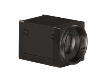
AcquCAM 23GR


More images with AcquCAM 23GR


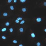
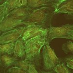
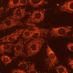
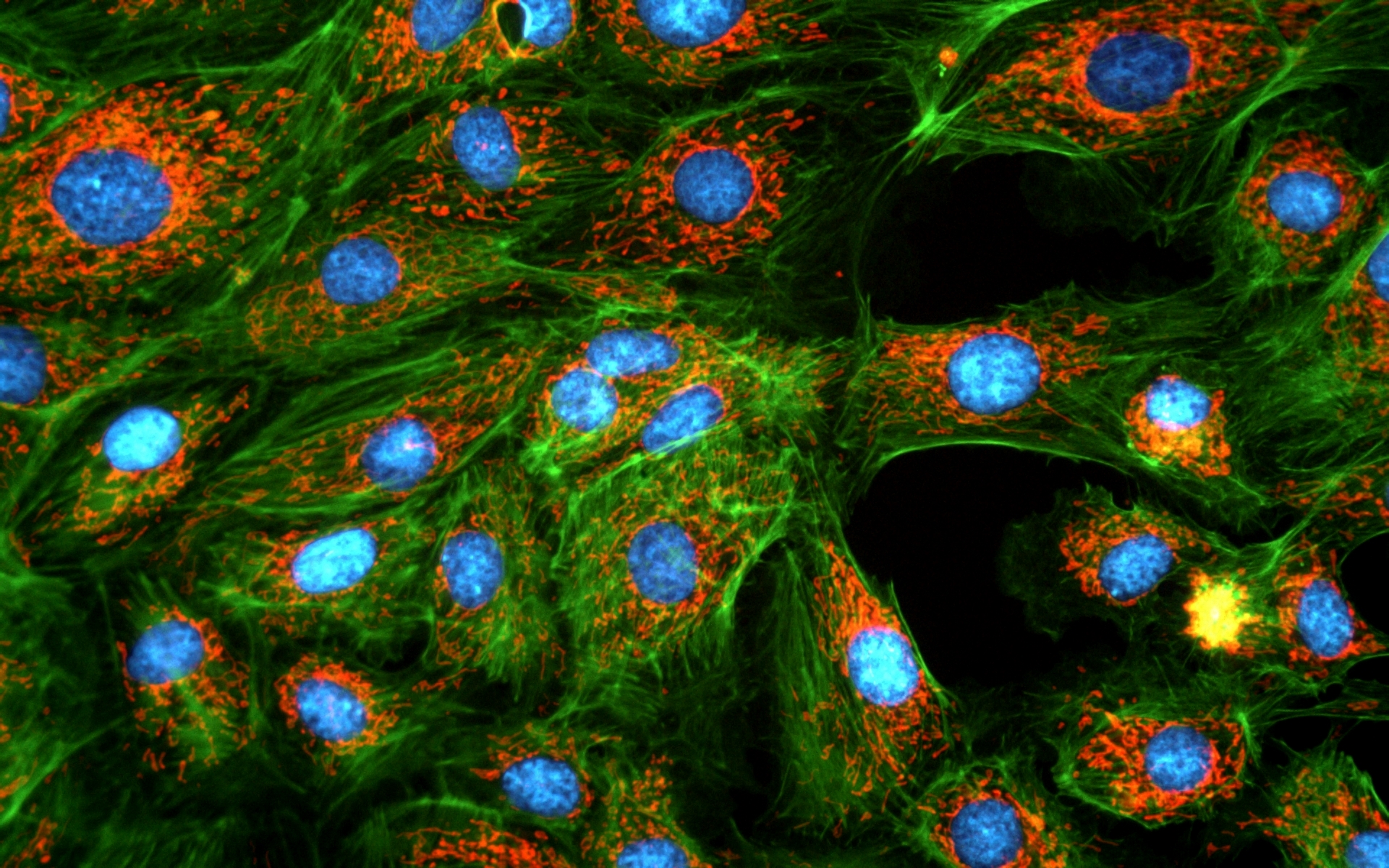
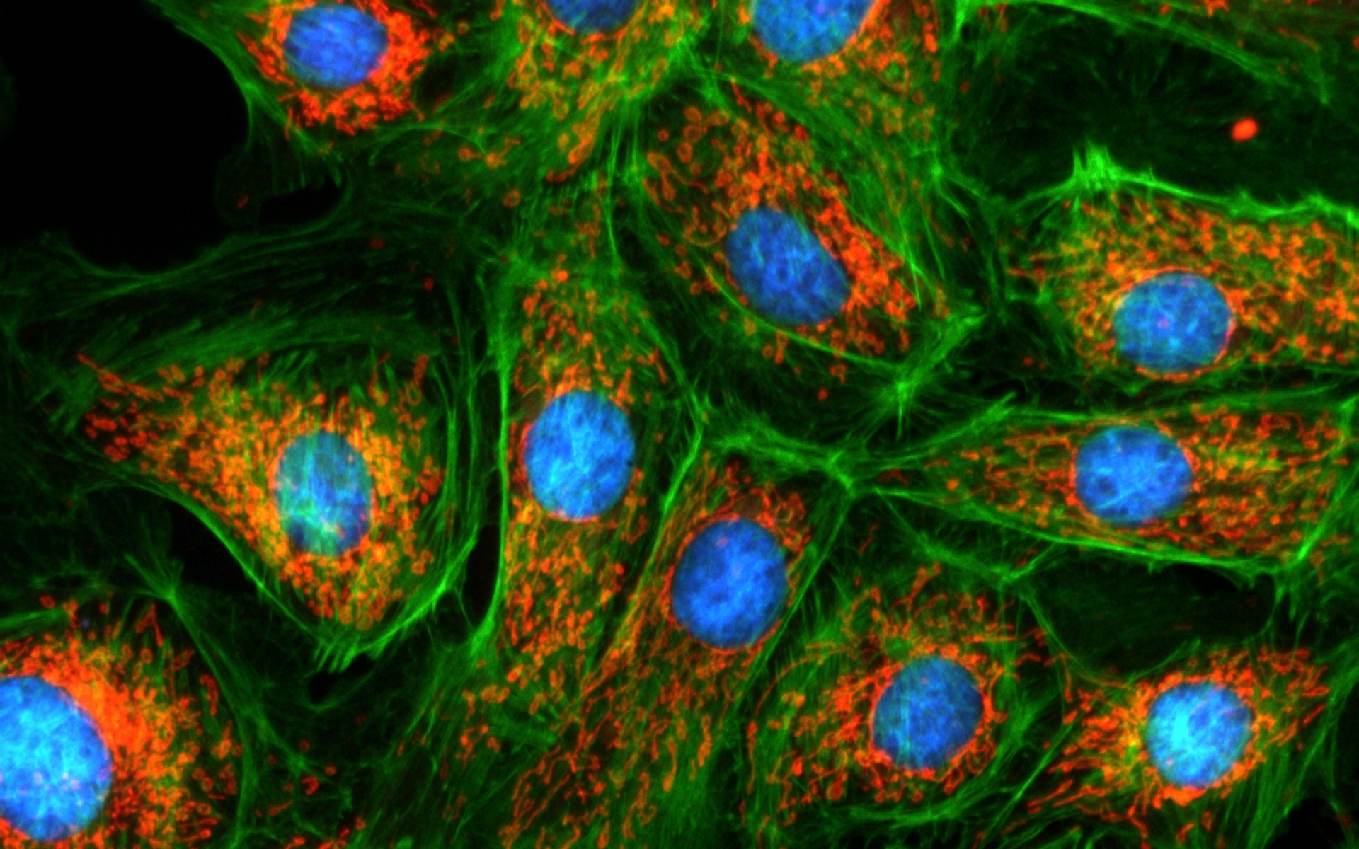
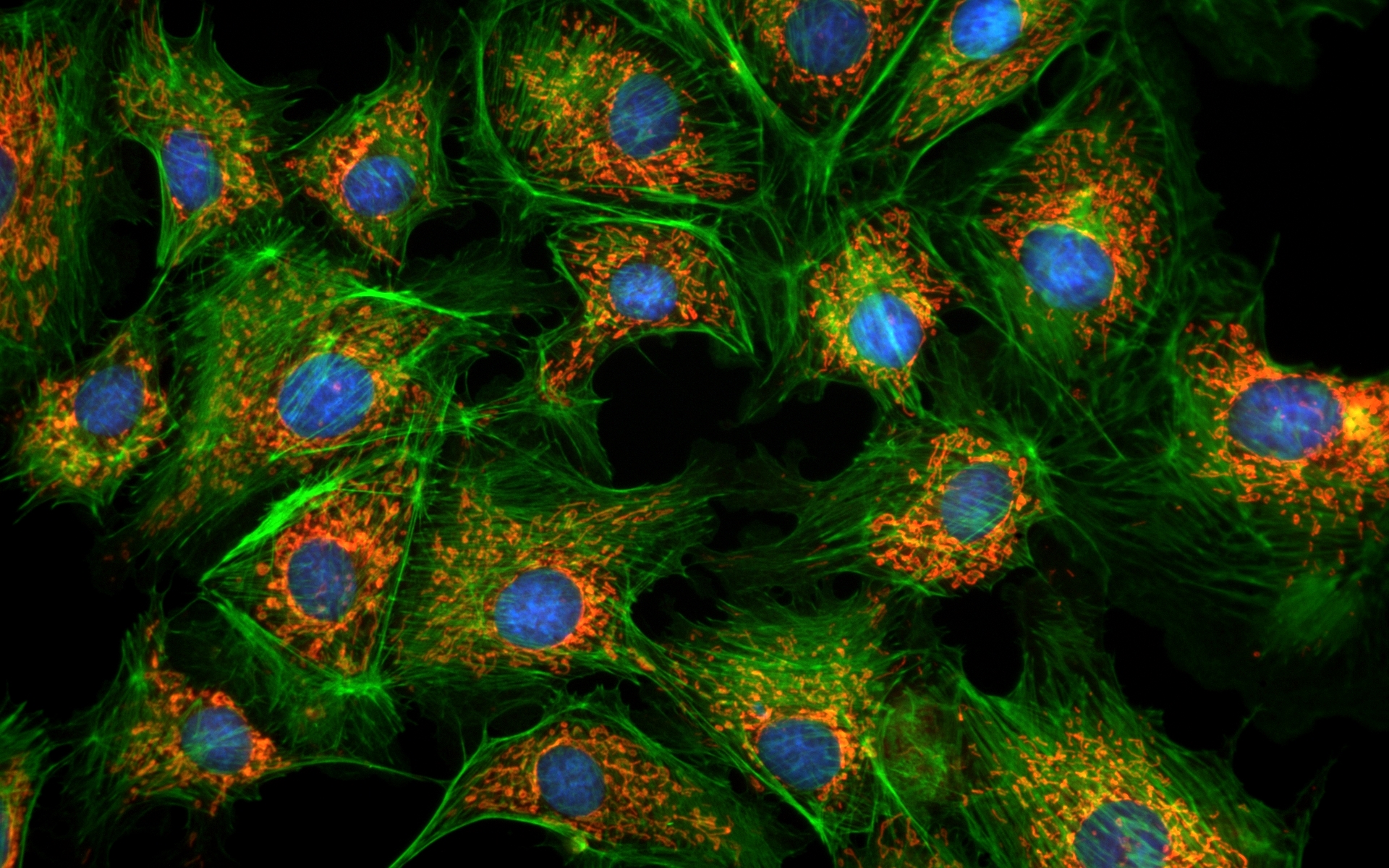
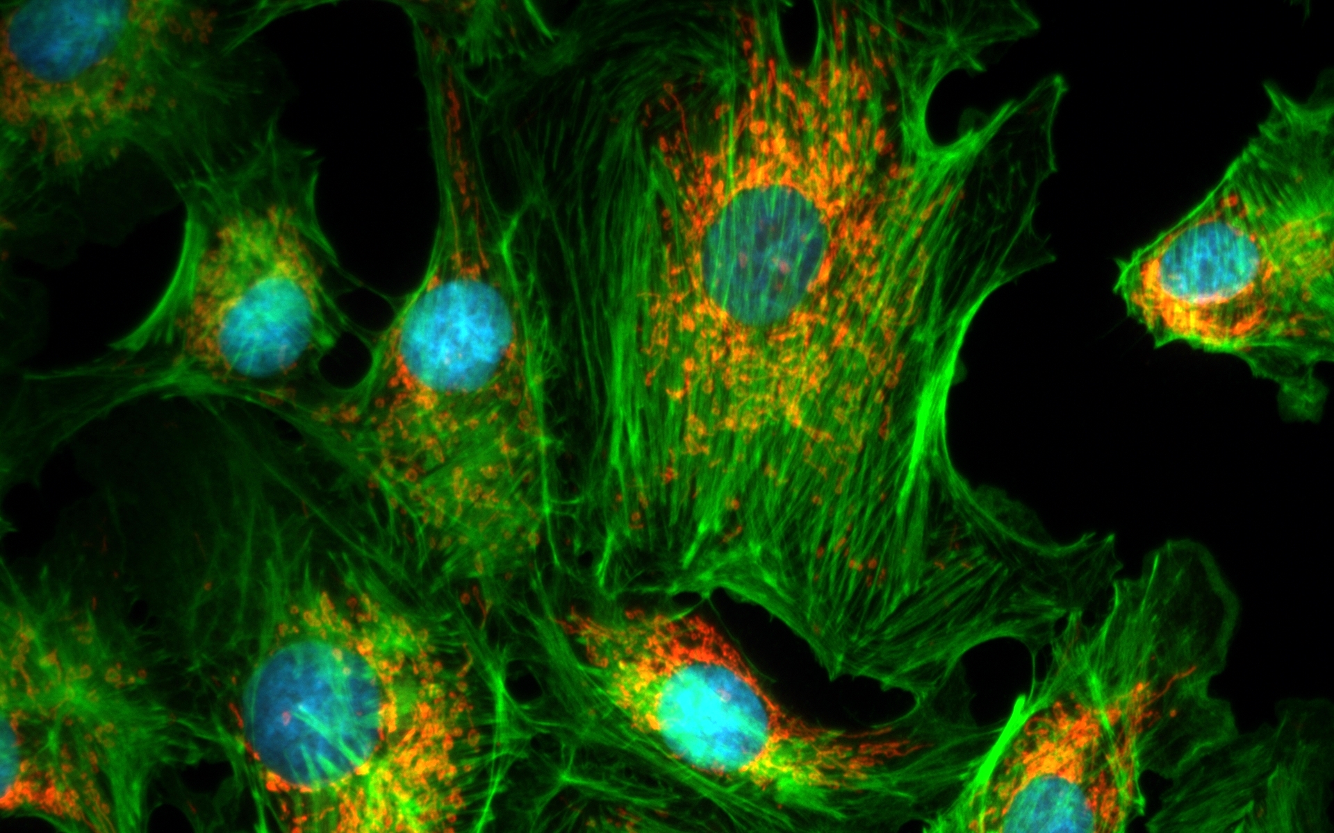
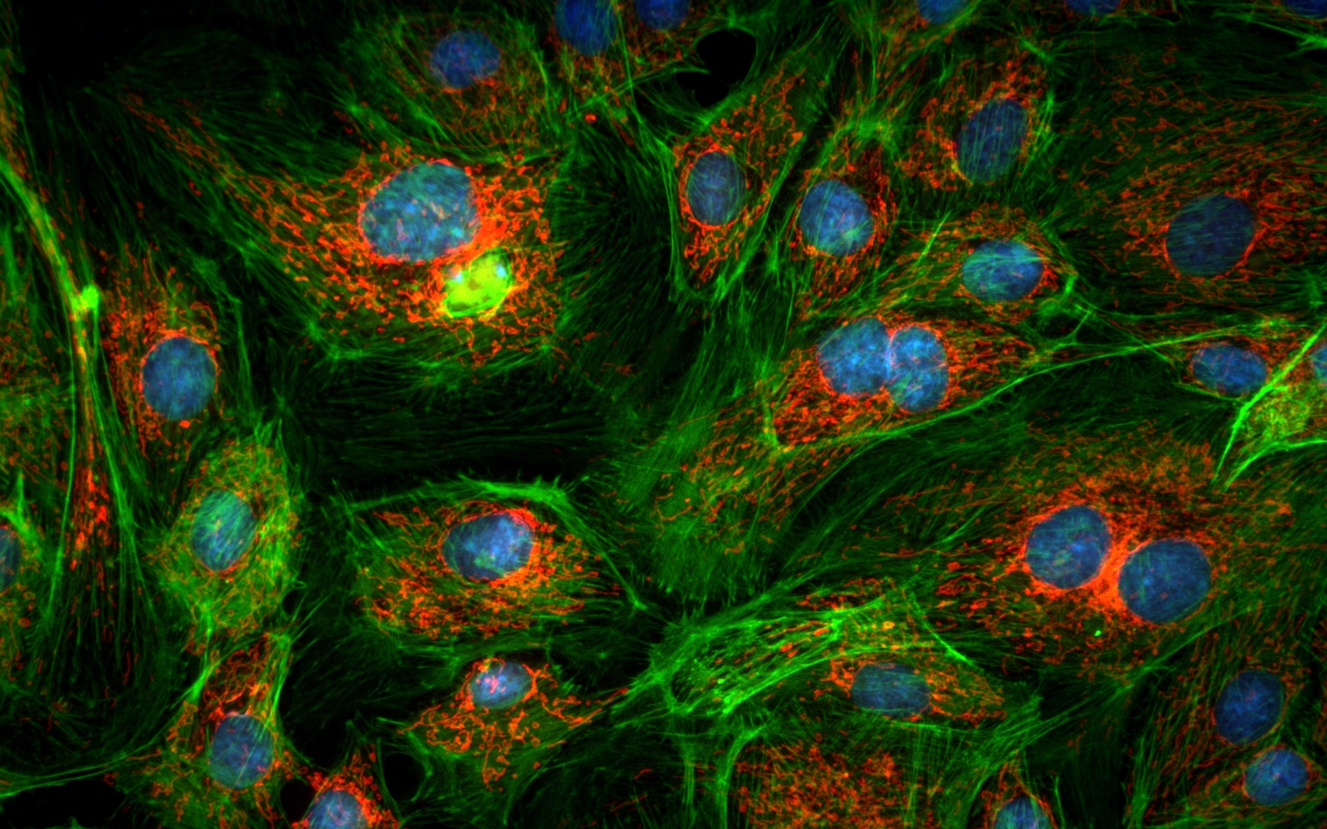
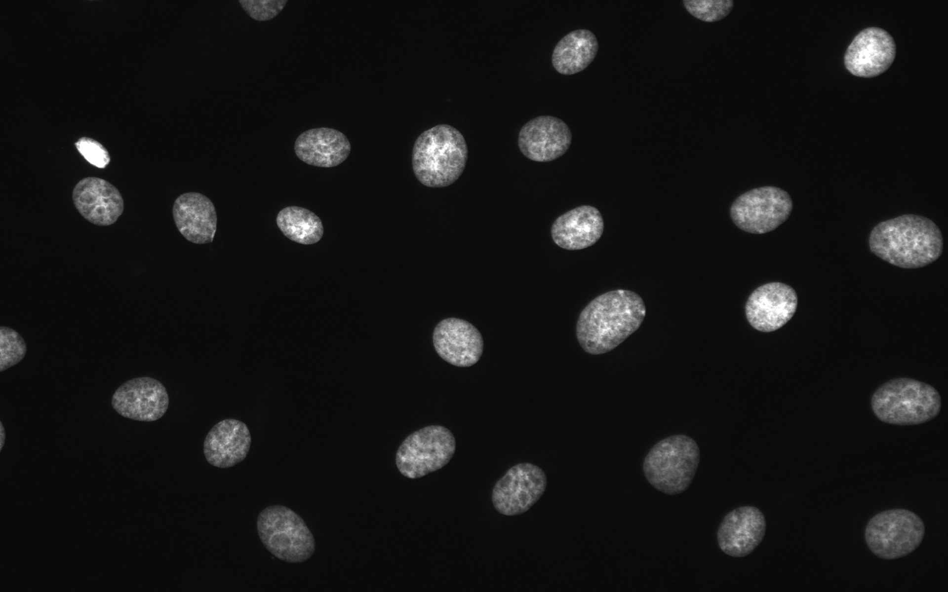
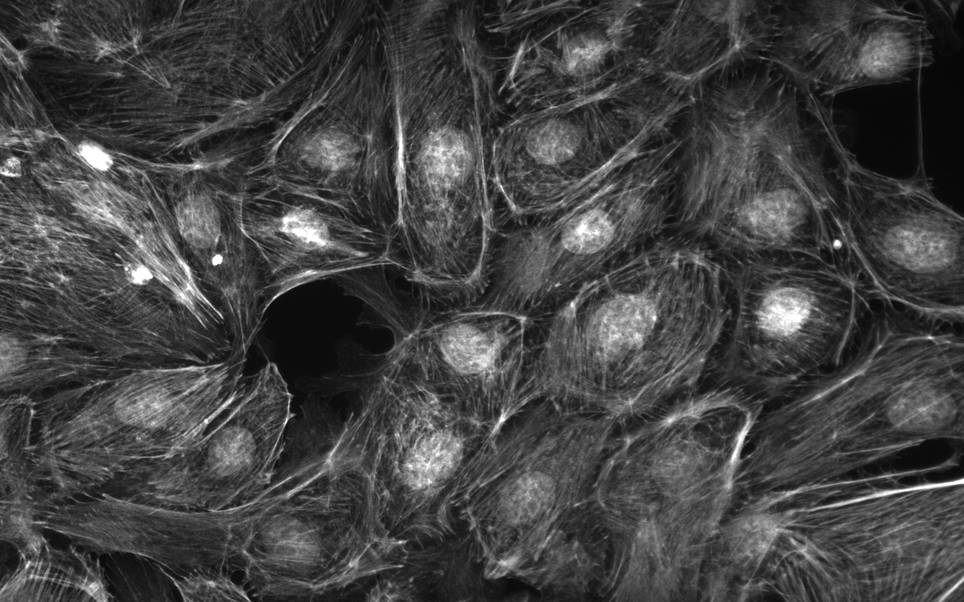
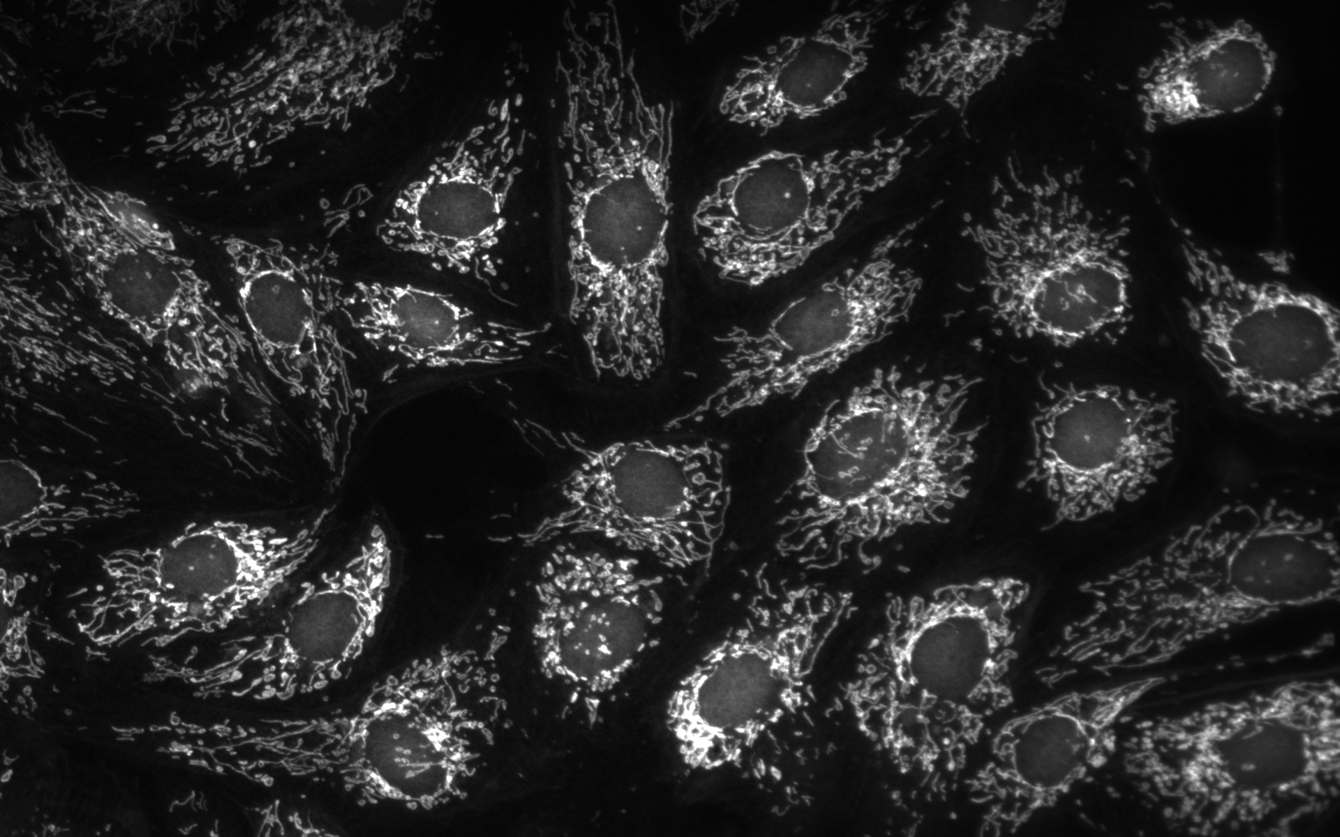
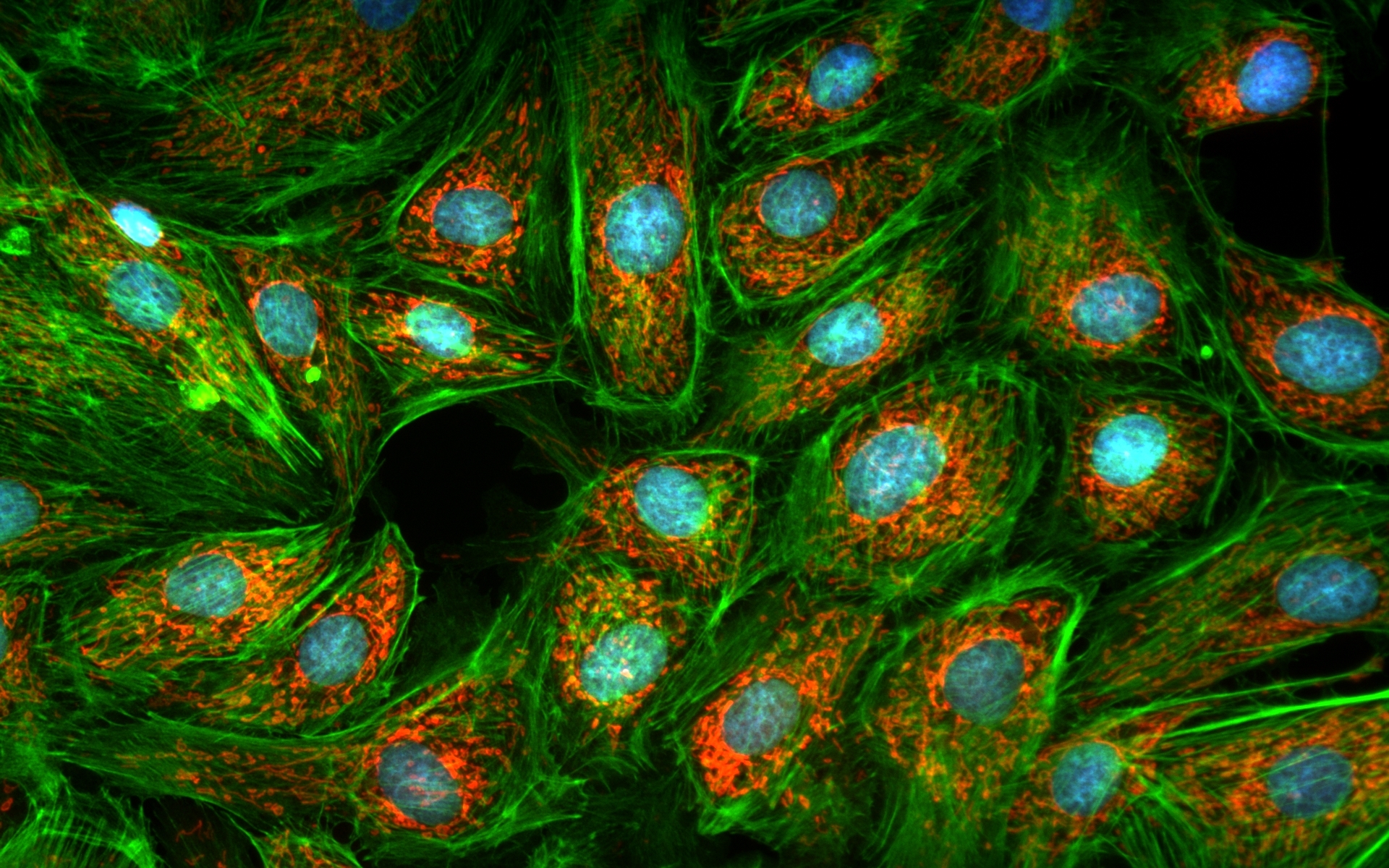
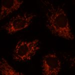
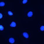
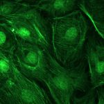
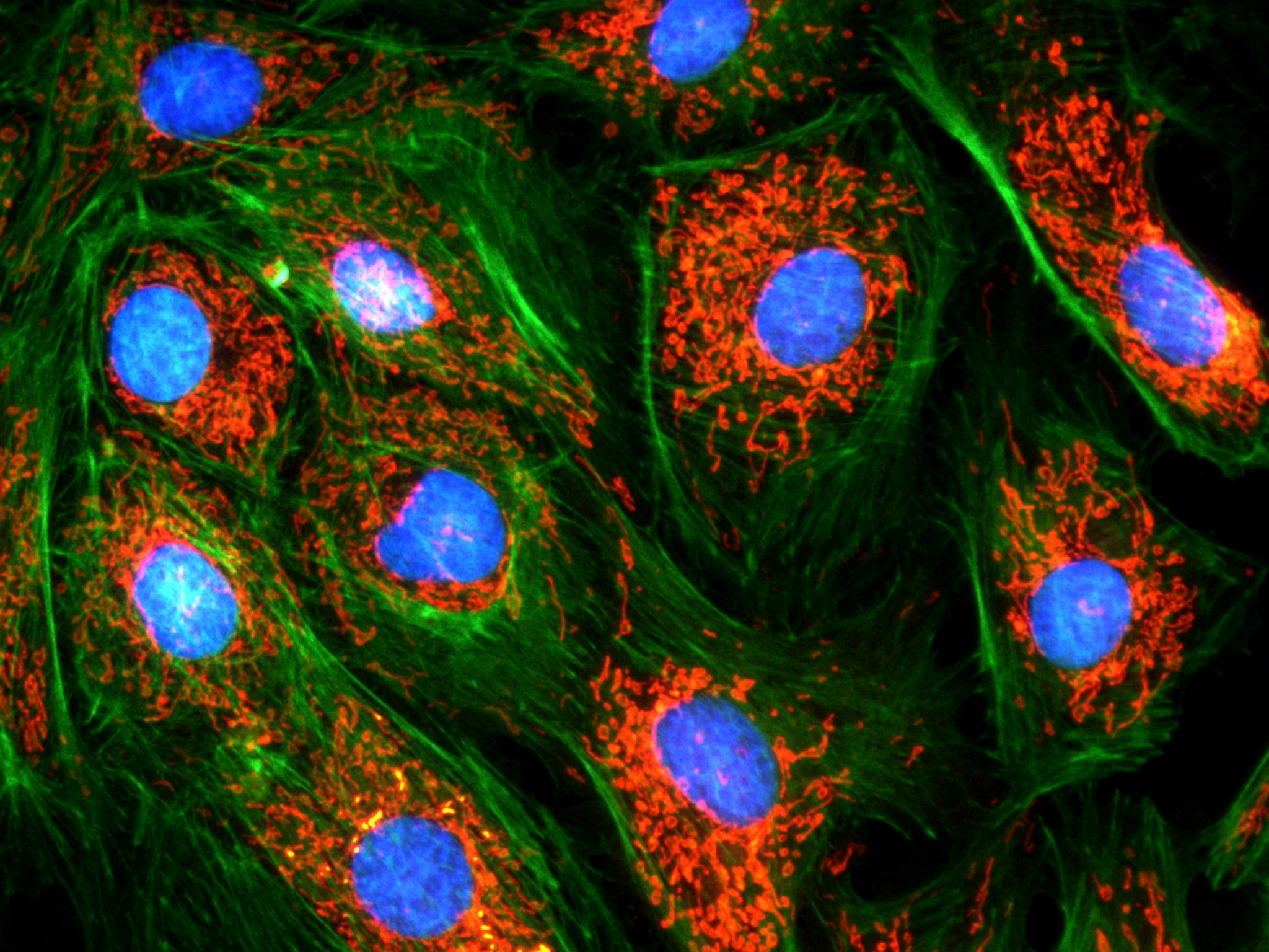
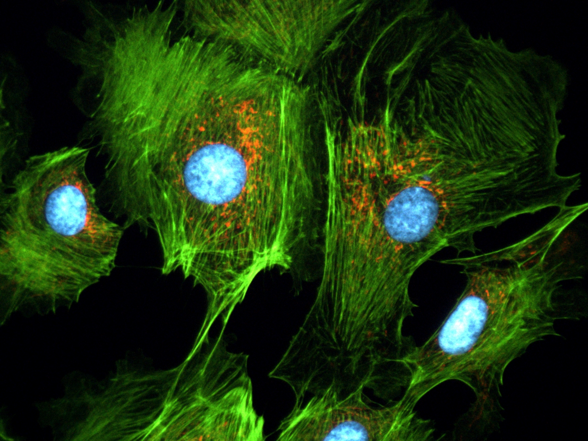
Sample : bovine pulmonary artery endothelial cell, BPAEC
Filter Set : JNO-U(L), B(L), G(L)
Camera : AcquCAM 23GR2
C-mount adapter : 1X
Lens : UPLNAPO40X
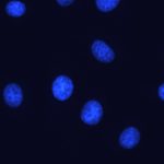
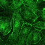
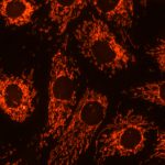
Above three images are merged and processed using ARM S/W


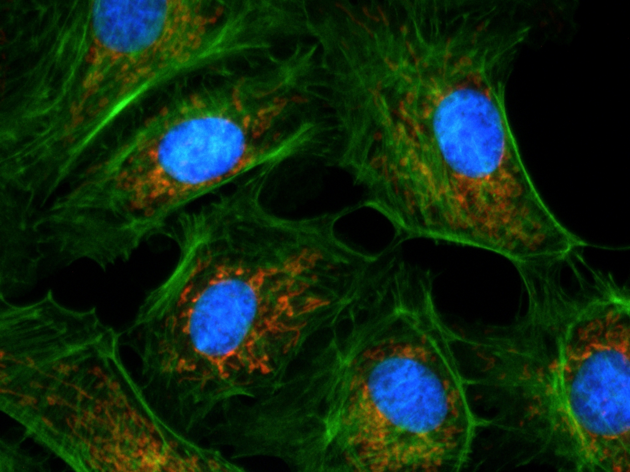
Sample : bovine pulmonary artery endothelial cell, BPAEC
Filter Set : JNO-U(B), B(B), G(B)
Camera : AcquCAM 23GR2
C-mount adapter : 1X
Lens : PlanApo60xWLSM
Above three images are merged and processed using ARM S/W


AcquCAM 23GR

AcquCAM 23GR


(Down load Catalog  – ACQUCAM 23GR)


  • USB 3.0 Color Camera
  • 1/1.2 ” Exmor Sensor
  • 1920×1200 pixel
  • Trigger input and I/O
  • Available  of taking images with a wavelength of 650nm or more, such as CY5
  • High Sensitivity Sensor (Best)
  • Fast, seamless image transfer
  • Available of images with a very wide field of view
  • Low Noise Sensor


The displays live digital images with gradual smoothness and combines exceptional resolution  with faithful color reproduction.The new sensors feature a global shutter function and able to capture a high-speed moving image without focal plane distortion. High-speed processing, low noise and low power dissipation by using column-parallel A/D conversion. equipped with trigger mode, and the external pulse can control accumulation time. The Sensor also have a pulse output function to indicate respective conditions during shutter operation and can be coordinated with peripheral circuits. High-definition images can be displayed live at a rate of high frames per second. without compression. Such imaging quality enables even the finest cellular regions to be observed clearly and distinctly without deterioration. While focusing is made stress free.

Images with AcquCAM 23GR


Sample : bovine pulmonary artery endothelial cell, BPAEC
Filter Set : JNO-U(B), B(B), G(B)
Camera : AcquCAM 23GR2
C-mount adapter : 1X
Lens : UPLNAPO40X
Above three images are merged and processed using ARM S/W


JNO-ARM

Advance Realtime Monitor. Easy-to-Use, Image Enhancement, etc


1. Information

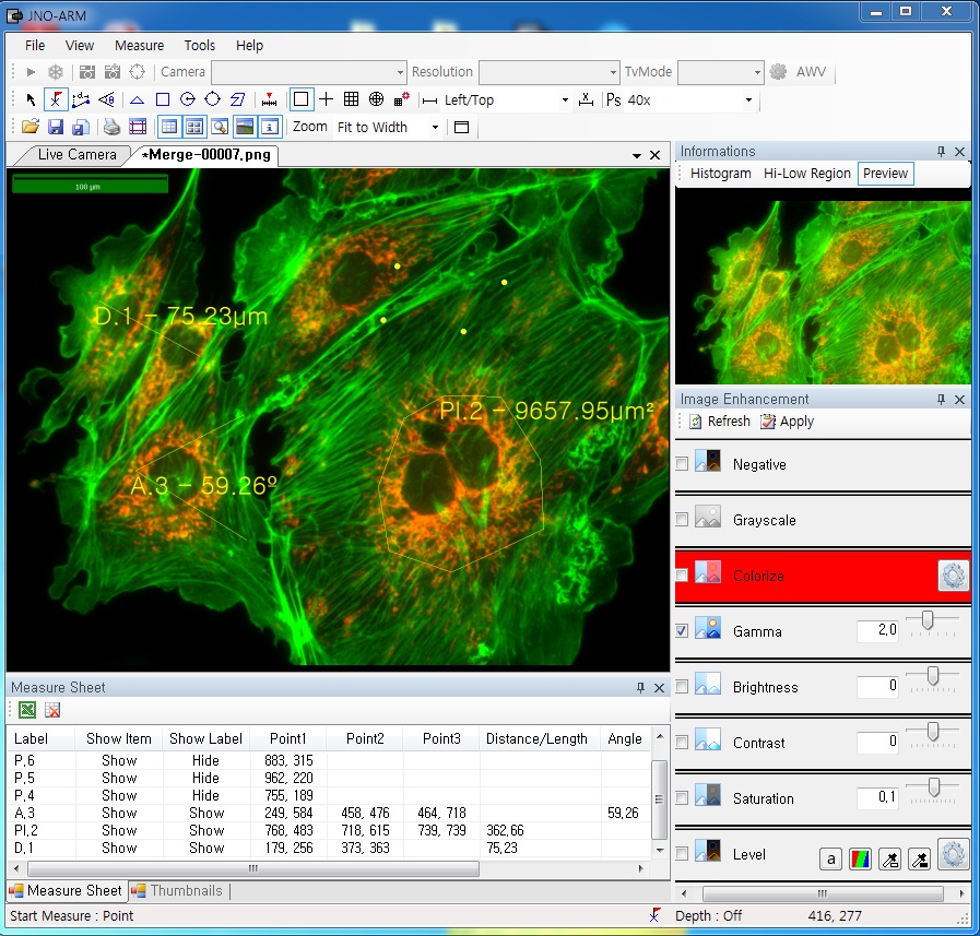
ARM is image analysis software for JNOPTIC AcquCAM cameras. This S/W is interchangeable with all WDM cameras regardless of camera brand and model and even more the most strength point is simple and easy use of length, area, angle, etc.


2. Simple measurement tools

  • Measurement Tools: Count, Distance, Angle, Area, etc.
  • Guide Lines: Cross Line, Rectangular Lattice, Circular Grid
  • Scale Panel on Live/Image Screen
  • Available measurement on LIVE image and saved image mode


3-1. Specialization for observation of fluorescence images

Improvement Function about Fluorescence Microscopy Image. (Used function: Auto Level, Low level, Pseudo Color, Merge Image)

Bottom on the left is merged image without improvement of image. And the bottom on the right is merged image after improvement of image. These images are shot by JNOPTIC AcquCAM Pro/G3 camera


3-2-1 Effect of high-contrast image with simple operation (Monochrome)

Improvement Function about Microscopy Image Monochrome. (Used function: Auto Level)

Effective improvement of image with simple operation. This image is shot by JNOPTIC Pro/S3 camera


3-2-2 Effect of high-contrast image with simple operation (Color)

Improvement Function about Microscopy Image. (Used function: Auto Level)

Effective improvement of image with simple operation. These images are shot by JNOPTIC AcquCAM Pro/G3 camera


3-3-1 Live Pseudo Color (for color camera)

When you get fluorescence images using color camera, if the unwanted channel was shown because of cross talk, you could remove this channel to take advantage of function of Live Pseudo.
Improvement Function about Fluorescence Microscopy Live Camera Image. (Used function: Live Pseudo Color)
 The extraction of wanted color information from color image (Remove BR on RGB source) These images are shot by JNOPTIC AcquCAM Pro/G3 camera

3-3-2 Live Pseudo Color (for Monochrome camera)

When you get fluorescence images using monochrome camera, you can raise efficiency of acquisition for fluorescence images to add similar false color to the color to be shown on the eyepiece in real time.
Improvement Function about Fluorescence Microscopy Live Monochrome Camera Image. (Used function: Live Pseudo Color)
 Improvement of usage environment for monochrome camera to add false color on the image. These images are shot by JNOPTIC AcquCAM Pro/S3 camera

4. JNO-MHU(Option Unit)

Available measuring height with additional unit, JNO-MHU

※ JNO-MHU is sold separately as optional unit.

Left: Focus on top of sample (Z-axis reset), Right: Focus on bottom of sample (Z-axis height measurement)

 < Measuring condition : over 20x objective, temp 20°C >

Responding Model Measurement value unit Measurable height Error of measuring 1000㎛ Recommended measuring height
CX 41 0.2 ㎛ -9.9 ~ 29.9㎜ Below ±20㎛ Below ± 2000㎛
CKX 41 0.2 ㎛ -9.9 ~ 29.9㎜ Below ±20㎛ Below ± 2000㎛
BX – FM 0.2 ㎛ -9.9 ~ 29.9㎜ Below ±20㎛ Below ± 2000㎛
BX 51/53 0.1 ㎛ -9.9 ~ 29.9㎜ Below ±10㎛ Below ± 1000㎛
MX 51 0.1 ㎛ -9.9 ~ 29.9㎜ Below ±10㎛ Below ± 1000㎛
MX 61L/61 0.1 ㎛ -9.9 ~ 29.9㎜ Below ±10㎛ Below ± 1000㎛


5. Reporting to Excel


Specification

Spec Description
Size 1/1.2 inch
Resolution H: 1920, V: 1200
Pixel size H: 5.86 µm, V: 5.86 µm
Video formats& Frame rate 1920×1200 RGB24@ 54 fps
1920×1200 Y800@ 162 fps
1920×1200 Y16@ 80 fps                             1920×1200 Y411 @ 108 fps
1920×1200 YUY2 @ 81 fps
Dynamic range 8 / 12 bit
Exposure time 20 ㎲ to 30s
Lens mount C/Cs mount
Interface USB 3.0
Power supply 4.5 to 5.5 VDC
Dimensions H: 29 mm, W: 29 mm, L: 43 mm
Mass 65g


More images with AcquCAM 23GR

-> For more details, click here.


AcquCAM 23S2 Images

AcquCAM 23S2


More images with AcquCAM 23S2


Sample : bovine pulmonary artery endothelial cell, BPAEC
Filter Set : JNO-U(L), B(L), G(L)
Camera : AcquCAM 23S2
C-mount adapter : 1X
Lens : UPLNAPO40X
Above three images are merged and processed using ARM S/W


Sample : bovine pulmonary artery endothelial cell, BPAEC
Filter Set : JNO-U(B), B(B), G(B)
Camera : AcquCAM 23S2
C-mount adapter : 1X
Lens : PlanApo60xWLSM
Above three images are merged and processed using ARM S/W


AcquCAM 23S2

AcquCAM 23S2

(Down load Catalog – AcquCAM 23S2)

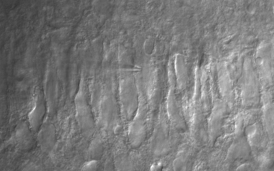
CA3 pyramidal neuron, IR-DIC & Alexa Fluo 488 at the same time
Figure courtesy by Dr. Sooyun Kim, Seoul National University
with Bx51WI, JNO-DPTS, AcquCAM 23S
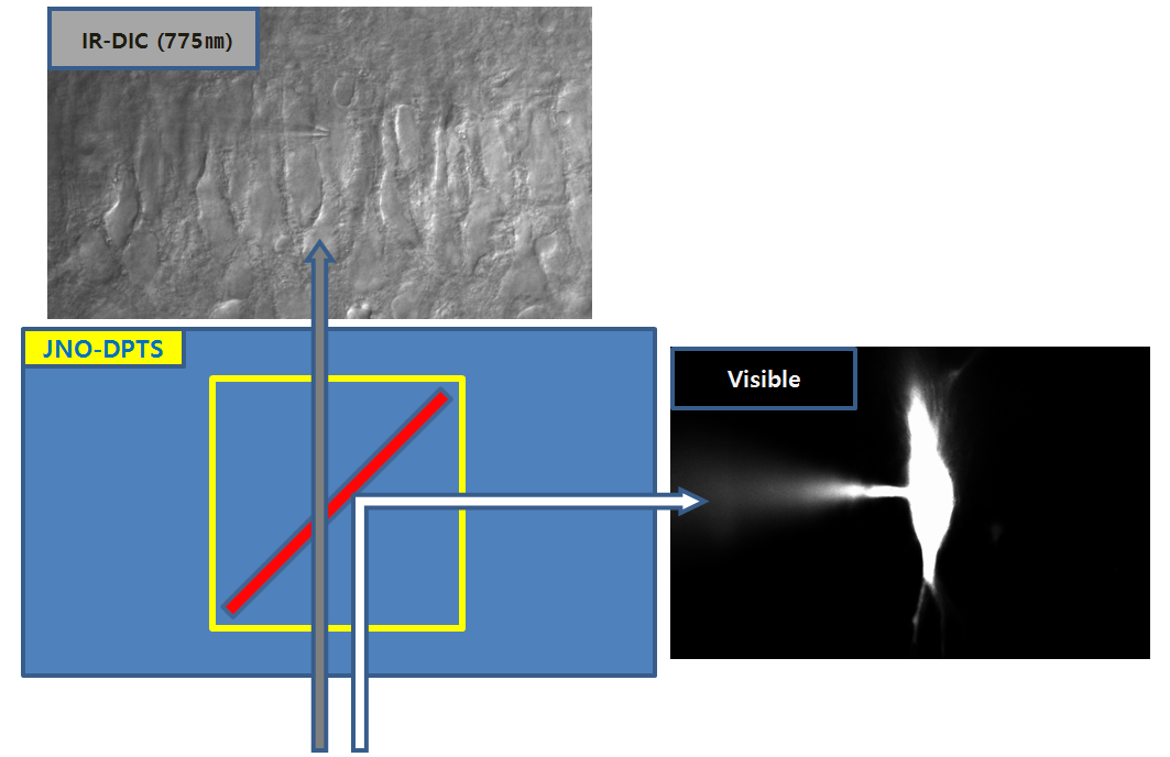
JNO-DPTS
CA3 pyramidal neuron, IR-DIC & Alexa Fluo 488 at the same time
Figure courtesy by Dr. Sooyun Kim, Seoul National University
with Bx51WI, JNO-DPTS, AcquCAM 23S
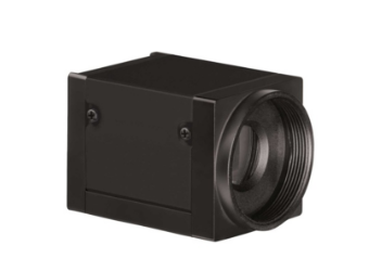
  • USB 3.0 Monochrome Camera
  • 1/1.2 ” Pregius Sensor
  • 1920×1200 pixel
  • Trigger input and I/O
  • Cost-effective mono camera for fluorescence microscope (Best)
  • Cost-effective camera for Department of Physiology (Best)
  • 1/1.2 inch optimized sensor size for microscope field of view
  • Acquire images of optimum width combined with 0.63x adapter

AcquCAM 23S2 mono camera has a USB 3.0 interface and is the perfect solution for many industrial automation, quality assurance, security, surveillance and medical applications. The mono camera ships with the very sensitive 1/1.2 inch CMOS Pregius  sensor. With up to 162 images per second, the AcuqCAM 23S2 is a low cost, yet highly versatile imaging solution. The camera includes a C to CS mount adapter, making it compatible to C and CS mount lenses.

Images with AcquCAM 23S2
Sample : bovine pulmonary artery endothelial cell, BPAEC
Filter Set : JNO-U(B), B(B), G(B)
Camera : AcquCAM 23S2
C-mount adapter : 1x
Lens : UPlanAPO 40x
Above three images are merged and processed using ARM S/W
Sample : bovine pulmonary artery endothelial cell, BPAEC
Filter Set : JNO-U(B), B(B), G(B)
Camera : AcquCAM 23S2
C-mount adapter : 1x
Lens : UPlanAPO 40x
Above three images are merged and processed using ARM S/W

Specification

SpecDescription
  Size  1/1.2 inch
  Resolution  H: 1920, V: 1200
 Pixel size  H: 5.86 µm, V: 5.86 µm
 Video formats & Frame rate1920×1200  Y16    @    80 fps
1920×1200  Y800 @ 162 fps
  Sensitivity  0.015 lx
  Exposure time  20㎲ to 30 s
  Lens mount  C/Cs mount
  Interface  USB 3.0
  Power supply  4.5 to 5.5 VDC
  Dimensions  H: 29 mm, W: 29 mm, L: 43 mm
  Mass  65 g

ARM

ARM is image analysis software for JNOPTIC AcquCAM cameras. This S/W is interchangeable with all WDM cameras regardless of camera brand and model and even more the most strength point is simple and easy use of length, area, angle, etc.

-> For more details, click here.

More images with AcquCAM 23S2

-> For more details, click here.

AcquCAM 3G Images

AcquCAM 3G


More images with AcquCAM 3G


Sample : bovine pulmonary artery endothelial cell, BPAEC
Filter Set : JNO-U(B), B(B), G(B)
Camera : AcquCAM 3G
C-mount adapter : 1X
Lens : PlanApo60xWLSM
Above three images are merged and processed using ARM S/W


Sample : bovine pulmonary artery endothelial cell, BPAEC
Filter Set : JNO-U(L), B(L), G(L)
Camera : AcquCAM 3G
C-mount adapter : 1X
Lens : UPLNAPO40X
Above three images are merged and processed using ARM S/W


AcquCAM 3G

AcquCAM 3G

Down load Catalog – ACQUCAM 3G

  • USB 3.0 Color Camera
  • 1/1.8 ” Pregius Sensor
  • 2048 x 1536 pixel
  • Trigger input and I/O
  • Cost-effective color camera
  • High Sensitivity Sensor
  • Fast, seamless image transfer
  • Available of images with a very wide field of view
  • Low Noise Sensor

AcquCAM 3G color camera has a USB 3.0 interface and is the perfect solution for many industrial automation, quality assurance, security, surveillance and medical applications. The color camera ships with the very sensitive 1/1.8 inch CMOS Pregius  sensor. With up to 60 images per second, the AcuqCAM 3G is a low cost, yet highly versatile imaging solution. The camera includes a C to CS mount adapter, making it compatible to C and CS mount lenses. Using the optional CS to M12 board lens adapter, the camera is
also compatible to M12 board lenses.

Images with AcquCAM 3G

Sample : bovine pulmonary artery endothelial cell, BPAEC
Filter Set : JNO-U(B), B(B), G(B)
Camera : AcquCAM 3G
C-mount adapter : 1X
Lens : UPLNAPO40X
Above three images are merged and processed using ARM S/W

Specification

Spec Description
Size 1/1.8 inch
Resolution H: 2048, V: 1536
Pixel size H: 3.45 µm, V: 3.45 µm
Video formats& Frame rate 2048×1536 (3.1 MP) RGB64 @ 60 fps

2048×1536 (3.1 MP) RGB32 @ 60 fps
2048×1536 (3.1 MP) Y16 @ 60 fps

2048×1536 (3.1 MP) Y411 @ 60 fps
2048×1536 (3.1 MP) YUY2 @ 60 fps

Dynamic range 8 / 12 bit
Exposure time 20 ㎲ to 30s
Lens mount C/Cs mount
Interface USB 3.0
Power supply 4.5 to 5.5 VDC
Dimensions H: 29 mm, W: 29 mm, L: 43 mm
Mass 65g

ARM

ARM is image analysis software for JNOPTIC AcquCAM cameras. This S/W is interchangeable with all WDM cameras regardless of camera brand and model and even more the most strength point is simple and easy use of length, area, angle, etc.

-> For more details, click here.

More images with AcquCAM 3G

-> For more details, click here.